1GJP | pdb_00001gjp

SCHIFF-BASE COMPLEX OF YEAST 5-AMINOLAEVULINIC ACID DEHYDRATASE WITH 4-OXOSEBACIC ACID


Experimental Data Snapshot

  • Method: X-RAY DIFFRACTION
  • Resolution: 1.80 Å
  • R-Value Free: 
    0.329 (Depositor) 
  • R-Value Work: 
    0.220 (DCC) 
  • R-Value Observed: 
    0.265 (Depositor) 

Starting Model: experimental
View more details

wwPDB Validation   3D Report Full Report


Ligand Structure Quality Assessment 

Created with Raphaël 2.3.0Worse 01 BetterLigand structure goodness of fit to experimental dataBest fitted 4OXClick on this verticalbar to view details

This is version 1.5 of the entry. See complete history


Literature

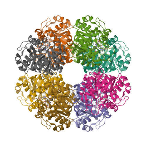
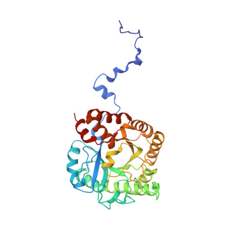
The X-Ray Structure of Yeast 5-Aminolaevulinic Acid Dehydratase Complexed with Two Diacid Inhibitors

Erskine, P.T.Coates, L.Newbold, R.Brindley, A.A.Stauffer, F.Wood, S.P.Warren, M.J.Cooper, J.B.Shoolingin-Jordan, P.M.Neier, R.

(2001) FEBS Lett 503: 196

  • DOI: https://doi.org/10.1016/s0014-5793(01)02721-1
  • Primary Citation of Related Structures:  
    1EB3, 1GJP

  • PubMed Abstract: 

    The structures of 5-aminolaevulinic acid dehydratase complexed with two irreversible inhibitors (4-oxosebacic acid and 4,7-dioxosebacic acid) have been solved at high resolution. Both inhibitors bind by forming a Schiff base link with Lys 263 at the active site. Previous inhibitor binding studies have defined the interactions made by only one of the two substrate moieties (P-side substrate) which bind to the enzyme during catalysis. The structures reported here provide an improved definition of the interactions made by both of the substrate molecules (A- and P-side substrates). The most intriguing result is the novel finding that 4,7-dioxosebacic acid forms a second Schiff base with the enzyme involving Lys 210. It has been known for many years that P-side substrate forms a Schiff base (with Lys 263) but until now there has been no evidence that binding of A-side substrate involves formation of a Schiff base with the enzyme. A catalytic mechanism involving substrate linked to the enzyme through Schiff bases at both the A- and P-sites is proposed.


  • Organizational Affiliation

    Division of Biochemistry and Molecular Biology, School of Biological Sciences, University of Southampton, UK.


Macromolecules
Find similar proteins by:  (by identity cutoff)  |  3D Structure
Entity ID: 1
MoleculeChains Sequence LengthOrganismDetailsImage
5-AMINOLAEVULINIC ACID DEHYDRATASE340Saccharomyces cerevisiaeMutation(s): 0 
EC: 4.2.1.24
UniProt
Find proteins for P05373 (Saccharomyces cerevisiae (strain ATCC 204508 / S288c))
Explore P05373 
Go to UniProtKB:  P05373
Entity Groups  
Sequence Clusters30% Identity50% Identity70% Identity90% Identity95% Identity100% Identity
UniProt GroupP05373
Sequence Annotations
Expand
  • Reference Sequence
Experimental Data & Validation

Experimental Data

  • Method: X-RAY DIFFRACTION
  • Resolution: 1.80 Å
  • R-Value Free:  0.329 (Depositor) 
  • R-Value Work:  0.220 (DCC) 
  • R-Value Observed: 0.265 (Depositor) 
Space Group: I 4 2 2
Unit Cell:
Length ( Å )Angle ( ˚ )
a = 103.5α = 90
b = 103.5β = 90
c = 168.2γ = 90
Software Package:
Software NamePurpose
SHELXL-97refinement
MOSFLMdata reduction
SCALAdata scaling
CCP4phasing

Structure Validation

View Full Validation Report


Ligand Structure Quality Assessment 

Created with Raphaël 2.3.0Worse 01 BetterLigand structure goodness of fit to experimental dataBest fitted 4OXClick on this verticalbar to view details

Entry History 

Deposition Data

Revision History  (Full details and data files)

  • Version 1.0: 2001-08-02
    Type: Initial release
  • Version 1.1: 2011-05-08
    Changes: Version format compliance
  • Version 1.2: 2011-07-13
    Changes: Version format compliance
  • Version 1.3: 2019-05-22
    Changes: Data collection, Derived calculations, Other, Refinement description
  • Version 1.4: 2023-12-13
    Changes: Data collection, Database references, Derived calculations, Other, Refinement description
  • Version 1.5: 2024-11-13
    Changes: Structure summary