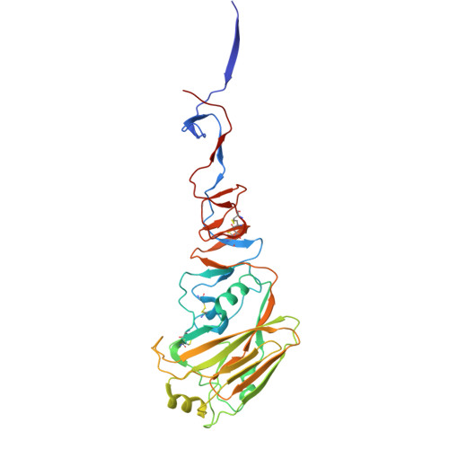
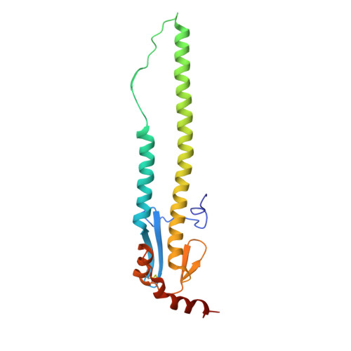
9DIP

Crystal structure of H5 hemagglutinin from the influenza virus A/Texas/37/2024 (H5N1) with LSTa


Experimental Data Snapshot

  • Method: X-RAY DIFFRACTION
  • Resolution: 2.32 Å
  • R-Value Free: 0.258 
  • R-Value Work: 0.216 
  • R-Value Observed: 0.218 

Starting Model: experimental
View more details

wwPDB Validation   3D Report Full Report


Ligand Structure Quality Assessment 


This is version 1.2 of the entry. See complete history


Literature

A single mutation in bovine influenza H5N1 hemagglutinin switches specificity to human receptors.

Lin, T.H.Zhu, X.Wang, S.Zhang, D.McBride, R.Yu, W.Babarinde, S.Paulson, J.C.Wilson, I.A.

(2024) Science 386: 1128-1134

  • DOI: https://doi.org/10.1126/science.adt0180
  • Primary Citation of Related Structures:  
    9DIO, 9DIP, 9DIQ

  • PubMed Abstract: 

    In 2024, several human infections with highly pathogenic clade 2.3.4.4b bovine influenza H5N1 viruses in the United States raised concerns about their capability for bovine-to-human or even human-to-human transmission. In this study, analysis of the hemagglutinin (HA) from the first-reported human-infecting bovine H5N1 virus (A/Texas/37/2024, Texas) revealed avian-type receptor binding preference. Notably, a Gln 226 Leu substitution switched Texas HA binding specificity to human-type receptors, which was enhanced when combined with an Asn 224 Lys mutation. Crystal structures of the Texas HA with avian receptor analog LSTa and its Gln 226 Leu mutant with human receptor analog LSTc elucidated the structural basis for this preferential receptor recognition. These findings highlight the need for continuous surveillance of emerging mutations in avian and bovine clade 2.3.4.4b H5N1 viruses.


  • Organizational Affiliation

    Department of Integrative Structural and Computational Biology, The Scripps Research Institute, La Jolla, CA, USA.


Macromolecules
Find similar proteins by:  (by identity cutoff)  |  3D Structure
Entity ID: 1
MoleculeChains Sequence LengthOrganismDetailsImage
Hemagglutinin HA1A,
C,
E [auth I]
325Influenza A virusMutation(s): 0 
Gene Names: HA
UniProt
Find proteins for A0A869KY97 (Influenza A virus)
Explore A0A869KY97 
Go to UniProtKB:  A0A869KY97
Entity Groups  
Sequence Clusters30% Identity50% Identity70% Identity90% Identity95% Identity100% Identity
UniProt GroupA0A869KY97
Glycosylation
Glycosylation Sites: 1
Sequence Annotations
Expand
  • Reference Sequence
Find similar proteins by:  (by identity cutoff)  |  3D Structure
Entity ID: 2
MoleculeChains Sequence LengthOrganismDetailsImage
Hemagglutinin HA2B,
D,
F [auth J]
176Influenza A virusMutation(s): 0 
Gene Names: HA
UniProt
Find proteins for A0A8K1XY81 (Influenza A virus)
Explore A0A8K1XY81 
Go to UniProtKB:  A0A8K1XY81
Entity Groups  
Sequence Clusters30% Identity50% Identity70% Identity90% Identity95% Identity100% Identity
UniProt GroupA0A8K1XY81
Sequence Annotations
Expand
  • Reference Sequence
Oligosaccharides

Help

Entity ID: 3
MoleculeChains Length2D Diagram Glycosylation3D Interactions
N-acetyl-alpha-neuraminic acid-(2-3)-beta-D-galactopyranose-(1-3)-2-acetamido-2-deoxy-beta-D-glucopyranose-(1-3)-beta-D-galactopyranoseG [auth E]4N/A
Glycosylation Resources
GlyTouCan:  G03309JT
GlyCosmos:  G03309JT
GlyGen:  G03309JT
Entity ID: 4
MoleculeChains Length2D Diagram Glycosylation3D Interactions
N-acetyl-alpha-neuraminic acid-(2-3)-beta-D-galactopyranose-(1-3)-2-acetamido-2-deoxy-beta-D-glucopyranose-(1-3)-beta-D-galactopyranose-(1-4)-alpha-D-glucopyranoseH [auth F],
J [auth H]
5N/A
Glycosylation Resources
GlyTouCan:  G08002FG
GlyCosmos:  G08002FG
Entity ID: 5
MoleculeChains Length2D Diagram Glycosylation3D Interactions
2-acetamido-2-deoxy-beta-D-glucopyranose-(1-4)-2-acetamido-2-deoxy-beta-D-glucopyranoseI [auth G]2N-Glycosylation
Glycosylation Resources
GlyTouCan:  G42666HT
GlyCosmos:  G42666HT
GlyGen:  G42666HT
Experimental Data & Validation

Experimental Data

  • Method: X-RAY DIFFRACTION
  • Resolution: 2.32 Å
  • R-Value Free: 0.258 
  • R-Value Work: 0.216 
  • R-Value Observed: 0.218 
  • Space Group: P 1 21 1
Unit Cell:
Length ( Å )Angle ( ˚ )
a = 103.539α = 90
b = 74.76β = 96.21
c = 131.101γ = 90
Software Package:
Software NamePurpose
PHENIXrefinement
HKL-3000data reduction
HKL-3000data scaling
PHASERphasing

Structure Validation

View Full Validation Report


Ligand Structure Quality Assessment 


Entry History & Funding Information

Deposition Data


Funding OrganizationLocationGrant Number
National Institutes of Health/National Institute Of Allergy and Infectious Diseases (NIH/NIAID)United States75N93021C00015

Revision History  (Full details and data files)

  • Version 1.0: 2024-12-04
    Type: Initial release
  • Version 1.1: 2024-12-11
    Changes: Structure summary
  • Version 1.2: 2024-12-18
    Changes: Database references