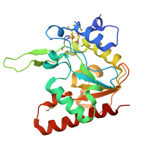
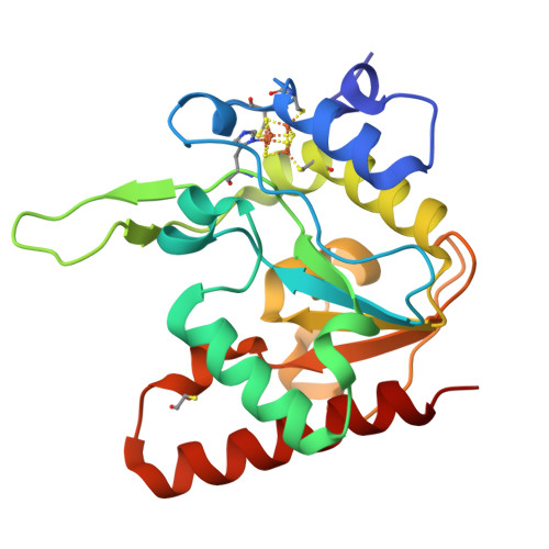
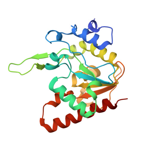
8IIM

H109K mutant of uracil DNA glycosylase X


Experimental Data Snapshot

  • Method: X-RAY DIFFRACTION
  • Resolution: 1.60 Å
  • R-Value Free: 0.182 
  • R-Value Work: 0.164 
  • R-Value Observed: 0.165 

Starting Model: experimental
View more details

wwPDB Validation   3D Report Full Report


Ligand Structure Quality Assessment 


This is version 1.2 of the entry. See complete history


Literature

Mutational and structural analyses of UdgX: insights into the active site pocket architecture and its evolution.

Aroli, S.Woo, E.J.Gopal, B.Varshney, U.

(2023) Nucleic Acids Res 51: 6554-6565

  • DOI: https://doi.org/10.1093/nar/gkad486
  • Primary Citation of Related Structures:  
    8IIE, 8IIF, 8IIG, 8IIH, 8III, 8IIJ, 8IIL, 8IIM, 8IIN, 8IIO, 8IIP, 8IIQ, 8IIR, 8IIS, 8IIT

  • PubMed Abstract: 

    UdgX excises uracil from uracil-containing DNA to concurrently form a covalent bond with the resulting AP-DNA. Structurally, UdgX is highly similar to family-4 UDGs (F4-UDGs). However, UdgX is unique in possessing a flexible R-loop (105KRRIH109). Among the class-defining motifs, while its motif A (51GEQPG55) diverged to possess Q53 in place of A53/G53 in F4-UDGs, motif B [178HPS(S/A)(L/V)(L/V)R184] has remained unchanged. Previously, we proposed an SN1 mechanism resulting in a covalent bond between H109 and AP-DNA. In this study, we investigated several single/double mutants of UdgX. The H109A, H109S, H109G, H109Q, H109C and H109K mutants gain conventional UDG activity to varying levels. The crystal structures of UdgX mutants show topological changes in their active sites, rationalizing their UDG activities. The E52Q, E52N and E52A mutants reveal that E52 forms a catalytic dyad with H109 to enhance its nucleophilicity. The Q53A mutant supports that UdgX specific evolution of Q53 occurred essentially to stabilize the R-loop conformation. The R184A mutation (motif B) supports the role of R184 in substrate-binding. Taken together, the structural, bioinformatics, and mutational studies suggest that UdgX diverged from F4-UDGs, and the emergence of the characteristic R-loop in UdgX is functionally assisted by A53/G53 to Q53 changes in motif A.


  • Organizational Affiliation

    Department of Microbiology and Cell Biology, Indian Institute of Science, Bangalore 560012, India.


Macromolecules
Find similar proteins by:  (by identity cutoff)  |  3D Structure
Entity ID: 1
MoleculeChains Sequence LengthOrganismDetailsImage
Type-4 uracil-DNA glycosylase208Mycolicibacterium smegmatis MC2 155Mutation(s): 1 
Gene Names: MSMEG_0265
EC: 3.2.2.27
UniProt
Find proteins for A0QP43 (Mycolicibacterium smegmatis (strain ATCC 700084 / mc(2)155))
Explore A0QP43 
Go to UniProtKB:  A0QP43
Entity Groups  
Sequence Clusters30% Identity50% Identity70% Identity90% Identity95% Identity100% Identity
UniProt GroupA0QP43
Sequence Annotations
Expand
  • Reference Sequence
Experimental Data & Validation

Experimental Data

  • Method: X-RAY DIFFRACTION
  • Resolution: 1.60 Å
  • R-Value Free: 0.182 
  • R-Value Work: 0.164 
  • R-Value Observed: 0.165 
  • Space Group: P 1 21 1
Unit Cell:
Length ( Å )Angle ( ˚ )
a = 36.45α = 90
b = 51.75β = 104.83
c = 54.63γ = 90
Software Package:
Software NamePurpose
PHENIXrefinement
Aimlessdata scaling
iMOSFLMdata reduction
PHASERphasing

Structure Validation

View Full Validation Report


Ligand Structure Quality Assessment 


Entry History & Funding Information

Deposition Data

  • Released Date: 2023-06-21 
  • Deposition Author(s): Aroli, S.

Funding OrganizationLocationGrant Number
Department of Biotechnology (DBT, India)IndiaBT/PR28058
Department of Biotechnology (DBT, India)IndiaBT/PR13522

Revision History  (Full details and data files)

  • Version 1.0: 2023-06-21
    Type: Initial release
  • Version 1.1: 2023-08-02
    Changes: Database references
  • Version 1.2: 2024-05-29
    Changes: Data collection