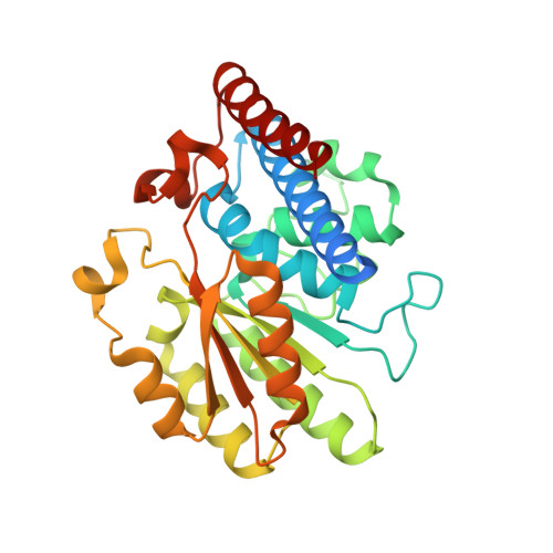
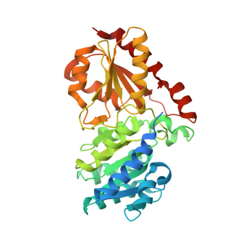
6YAK

Split gene transketolase, active alpha2beta2 heterotetramer


Experimental Data Snapshot

  • Method: X-RAY DIFFRACTION
  • Resolution: 1.34 Å
  • R-Value Free: 0.159 
  • R-Value Work: 0.141 

Starting Model: experimental
View more details

wwPDB Validation   3D Report Full Report


Ligand Structure Quality Assessment 


This is version 1.1 of the entry. See complete history


Literature

A 'Split-Gene' Transketolase From the Hyper-Thermophilic Bacterium Carboxydothermus hydrogenoformans : Structure and Biochemical Characterization.

James, P.Isupov, M.N.De Rose, S.A.Sayer, C.Cole, I.S.Littlechild, J.A.

(2020) Front Microbiol 11: 592353-592353

  • DOI: https://doi.org/10.3389/fmicb.2020.592353
  • Primary Citation of Related Structures:  
    6YAJ, 6YAK

  • PubMed Abstract: 

    A novel transketolase has been reconstituted from two separate polypeptide chains encoded by a 'split-gene' identified in the genome of the hyperthermophilic bacterium, Carboxydothermus hydrogenoformans . The reconstituted active α 2 β 2 tetrameric enzyme has been biochemically characterized and its activity has been determined using a range of aldehydes including glycolaldehyde, phenylacetaldehyde and cyclohexanecarboxaldehyde as the ketol acceptor and hydroxypyruvate as the donor. This reaction proceeds to near 100% completion due to the release of the product carbon dioxide and can be used for the synthesis of a range of sugars of interest to the pharmaceutical industry. This novel reconstituted transketolase is thermally stable with no loss of activity after incubation for 1 h at 70°C and is stable after 1 h incubation with 50% of the organic solvents methanol, ethanol, isopropanol, DMSO, acetonitrile and acetone. The X-ray structure of the holo reconstituted α 2 β 2 tetrameric transketolase has been determined to 1.4 Å resolution. In addition, the structure of an inactive tetrameric β 4 protein has been determined to 1.9 Å resolution. The structure of the active reconstituted α 2 β 2 enzyme has been compared to the structures of related enzymes; the E1 component of the pyruvate dehydrogenase complex and D-xylulose-5-phosphate synthase, in an attempt to rationalize differences in structure and substrate specificity between these enzymes. This is the first example of a reconstituted 'split-gene' transketolase to be biochemically and structurally characterized allowing its potential for industrial biocatalysis to be evaluated.


  • Organizational Affiliation

    Henry Wellcome Building for Biocatalysis, Biosciences, University of Exeter, Exeter, United Kingdom.


Macromolecules
Find similar proteins by:  (by identity cutoff)  |  3D Structure
Entity ID: 1
MoleculeChains Sequence LengthOrganismDetailsImage
N-terminal component of the split chain transketolaseA [auth AAA],
C [auth CCC]
309Carboxydothermus hydrogenoformansMutation(s): 0 
Entity Groups  
Sequence Clusters30% Identity50% Identity70% Identity90% Identity95% Identity100% Identity
Sequence Annotations
Expand
  • Reference Sequence
Find similar proteins by:  (by identity cutoff)  |  3D Structure
Entity ID: 2
MoleculeChains Sequence LengthOrganismDetailsImage
C-terminal component of the split chain transketolaseB [auth BBB],
D [auth DDD]
341Carboxydothermus hydrogenoformansMutation(s): 0 
Entity Groups  
Sequence Clusters30% Identity50% Identity70% Identity90% Identity95% Identity100% Identity
Sequence Annotations
Expand
  • Reference Sequence
Small Molecules
Ligands 5 Unique
IDChains Name / Formula / InChI Key2D Diagram3D Interactions
8EL (Subject of Investigation/LOI)
Query on 8EL

Download Ideal Coordinates CCD File 
E [auth AAA],
S [auth CCC]
2-[3-[(4-azanyl-2-methyl-pyrimidin-5-yl)methyl]-4-methyl-2H-1,3-thiazol-5-yl]ethyl phosphono hydrogen phosphate
C12 H20 N4 O7 P2 S
FCUXBJDJFWOWMD-UHFFFAOYSA-N
MLT
Query on MLT

Download Ideal Coordinates CCD File 
H [auth AAA]
I [auth AAA]
N [auth BBB]
O [auth BBB]
P [auth BBB]
H [auth AAA],
I [auth AAA],
N [auth BBB],
O [auth BBB],
P [auth BBB],
Q [auth BBB],
V [auth CCC],
W [auth CCC],
X [auth CCC],
Y [auth CCC],
Z [auth CCC]
D-MALATE
C4 H6 O5
BJEPYKJPYRNKOW-UWTATZPHSA-N
LMR
Query on LMR

Download Ideal Coordinates CCD File 
BA [auth DDD],
CA [auth DDD],
J [auth AAA],
K [auth AAA],
L [auth AAA]
(2S)-2-hydroxybutanedioic acid
C4 H6 O5
BJEPYKJPYRNKOW-REOHCLBHSA-N
CA (Subject of Investigation/LOI)
Query on CA

Download Ideal Coordinates CCD File 
F [auth AAA],
G [auth AAA],
T [auth CCC],
U [auth CCC]
CALCIUM ION
Ca
BHPQYMZQTOCNFJ-UHFFFAOYSA-N
CL
Query on CL

Download Ideal Coordinates CCD File 
AA [auth CCC],
DA [auth DDD],
M [auth AAA],
R [auth BBB]
CHLORIDE ION
Cl
VEXZGXHMUGYJMC-UHFFFAOYSA-M
Experimental Data & Validation

Experimental Data

Unit Cell:
Length ( Å )Angle ( ˚ )
a = 123.078α = 90
b = 130.047β = 90
c = 165.897γ = 90
Software Package:
Software NamePurpose
REFMACrefinement
XDSdata reduction
XSCALEdata scaling
Aimlessdata scaling
MOLREPphasing
MoRDaphasing

Structure Validation

View Full Validation Report


Ligand Structure Quality Assessment 


Entry History & Funding Information

Deposition Data


Funding OrganizationLocationGrant Number
Biotechnology and Biological Sciences Research Council (BBSRC)United KingdomBB/L002035/1

Revision History  (Full details and data files)

  • Version 1.0: 2020-11-25
    Type: Initial release
  • Version 1.1: 2024-01-24
    Changes: Data collection, Database references, Derived calculations, Refinement description