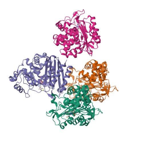
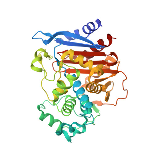
RCSB PDB - 6TZH: ADC-7 in complex with boronic acid transition state inhibitor S06015

 6TZH

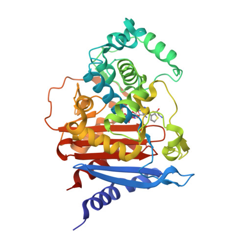
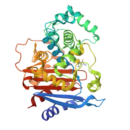
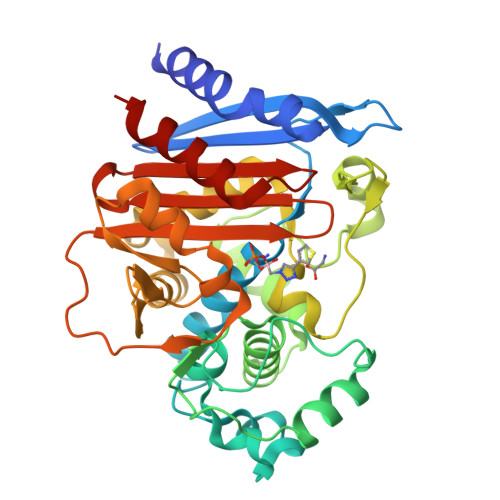
ADC-7 in complex with boronic acid transition state inhibitor S06015


Experimental Data Snapshot

  • Method: X-RAY DIFFRACTION
  • Resolution: 2.04 Å
  • R-Value Free: 0.254 
  • R-Value Work: 0.221 
  • R-Value Observed: 0.223 

wwPDB Validation   3D Report Full Report


Ligand Structure Quality Assessment 

Created with Raphaël 2.3.0Worse 01 BetterLigand structure goodness of fit to experimental dataBest fitted ERFClick on this verticalbar to view details

This is version 1.3 of the entry. See complete history


Literature

1,2,3-Triazolylmethaneboronate: A Structure Activity Relationship Study of a Class of beta-Lactamase Inhibitors againstAcinetobacter baumanniiCephalosporinase.

Caselli, E.Fini, F.Introvigne, M.L.Stucchi, M.Taracila, M.A.Fish, E.R.Smolen, K.A.Rather, P.N.Powers, R.A.Wallar, B.J.Bonomo, R.A.Prati, F.

(2020) ACS Infect Dis 6: 1965-1975

  • DOI: https://doi.org/10.1021/acsinfecdis.0c00254
  • Primary Citation of Related Structures:  
    6TZF, 6TZG, 6TZH, 6TZI, 6TZJ

  • PubMed Abstract: 

    Boronic acid transition state inhibitors (BATSIs) are known reversible covalent inhibitors of serine β-lactamases. The selectivity and high potency of specific BATSIs bearing an amide side chain mimicking the β-lactam's amide side chain are an established and recognized synthetic strategy. Herein, we describe a new class of BATSIs where the amide group is replaced by a bioisostere triazole; these compounds were designed as molecular probes. To this end, a library of 26 α-triazolylmethaneboronic acids was synthesized and tested against the clinically concerning Acinetobacter -derived cephalosporinase, ADC-7. In steady state analyses, these compounds demonstrated K i values ranging from 90 nM to 38 μM (±10%). Five compounds were crystallized in complex with ADC-7 β-lactamase, and all the crystal structures reveal the triazole is in the putative amide binding site, thus confirming the triazole-amide bioisosterism. The easy synthetic access of these new inhibitors as prototype scaffolds allows the insertion of a wide range of chemical groups able to explore the enzyme binding site and provides insights on the importance of specific residues in recognition and catalysis. The best inhibitor identified, compound 6q ( K i 90 nM), places a tolyl group near Arg340, making favorable cation-π interactions. Notably, the structure of 6q does not resemble the natural substrate of the β-lactamase yet displays a pronounced inhibition activity, in addition to lowering the minimum inhibitory concentration (MIC) of ceftazidime against three bacterial strains expressing class C β-lactamases. In summary, these observations validate the α-triazolylboronic acids as a promising template for further inhibitor design.


  • Organizational Affiliation

    Department of Life Sciences, University of Modena and Reggio Emilia, via Campi 103, Modena 41125, Italy.


Macromolecules
Find similar proteins by:  (by identity cutoff)  |  3D Structure
Entity ID: 1
MoleculeChains Sequence LengthOrganismDetailsImage
Beta-lactamase
A, B, C, D
361Acinetobacter baumanniiMutation(s): 0 
EC: 3.5.2.6
UniProt
Find proteins for Q6DRA1 (Acinetobacter baumannii)
Explore Q6DRA1 
Go to UniProtKB:  Q6DRA1
Entity Groups  
Sequence Clusters30% Identity50% Identity70% Identity90% Identity95% Identity100% Identity
UniProt GroupQ6DRA1
Sequence Annotations
Expand
  • Reference Sequence
Small Molecules
Ligands 3 Unique
IDChains Name / Formula / InChI Key2D Diagram3D Interactions
ERF (Subject of Investigation/LOI)
Query on ERF

Download Ideal Coordinates CCD File 
E [auth A],
G [auth B],
K [auth C],
M [auth D]
phosphonooxy-[(4-thiophen-3-yl-1,2,3-triazol-1-yl)methyl]borinic acid
C7 H9 B N3 O5 P S
CSAHOMHDQOWLFN-UHFFFAOYSA-N
PO4
Query on PO4

Download Ideal Coordinates CCD File 
H [auth B]PHOSPHATE ION
O4 P
NBIIXXVUZAFLBC-UHFFFAOYSA-K
GLY
Query on GLY

Download Ideal Coordinates CCD File 
F [auth A],
I [auth B],
J [auth B],
L [auth C],
N [auth D]
GLYCINE
C2 H5 N O2
DHMQDGOQFOQNFH-UHFFFAOYSA-N
Experimental Data & Validation

Experimental Data

  • Method: X-RAY DIFFRACTION
  • Resolution: 2.04 Å
  • R-Value Free: 0.254 
  • R-Value Work: 0.221 
  • R-Value Observed: 0.223 
  • Space Group: P 1 21 1
Unit Cell:
Length ( Å )Angle ( ˚ )
a = 88.554α = 90
b = 81.464β = 113.1
c = 105.665γ = 90
Software Package:
Software NamePurpose
REFMACrefinement
PDB_EXTRACTdata extraction
XDSdata reduction
Aimlessdata scaling
PHASERphasing
Cootmodel building

Structure Validation

View Full Validation Report


Ligand Structure Quality Assessment 

Created with Raphaël 2.3.0Worse 01 BetterLigand structure goodness of fit to experimental dataBest fitted ERFClick on this verticalbar to view details

Entry History & Funding Information

Deposition Data


Funding OrganizationLocationGrant Number
National Institutes of Health/National Institute Of Allergy and Infectious Diseases (NIH/NIAID)United StatesR01 AI072219

Revision History  (Full details and data files)

  • Version 1.0: 2020-06-24
    Type: Initial release
  • Version 1.1: 2020-07-08
    Changes: Database references
  • Version 1.2: 2020-07-22
    Changes: Database references
  • Version 1.3: 2024-10-23
    Changes: Data collection, Database references, Refinement description, Structure summary