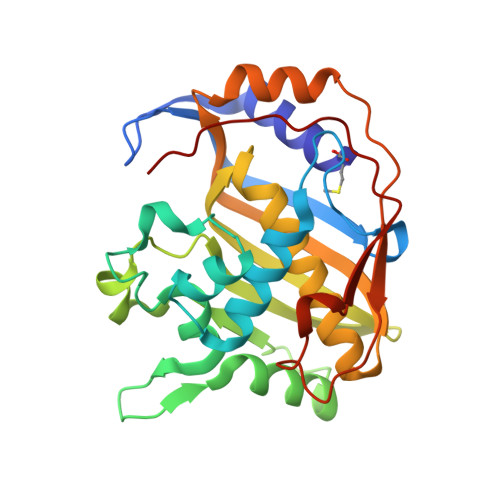
6NNR

high-resolution structure of wild-type E. coli thymidylate synthase


Experimental Data Snapshot

  • Method: X-RAY DIFFRACTION
  • Resolution: 1.05 Å
  • R-Value Free: 0.132 
  • R-Value Work: 0.121 
  • R-Value Observed: 0.121 

Starting Model: experimental
View more details

wwPDB Validation   3D Report Full Report


Ligand Structure Quality Assessment 


This is version 1.3 of the entry. See complete history


Literature

Mg2+ binds to the surface of thymidylate synthase and affects hydride transfer at the interior active site.

Wang, Z.Sapienza, P.J.Abeysinghe, T.Luzum, C.Lee, A.L.Finer-Moore, J.S.Stroud, R.M.Kohen, A.

(2013) J Am Chem Soc 135: 7583-7592

  • DOI: https://doi.org/10.1021/ja400761x
  • Primary Citation of Related Structures:  
    6NNR

  • PubMed Abstract: 

    Thymidylate synthase (TSase) produces the sole intracellular de novo source of thymidine (i.e., the DNA base T) and thus is a common target for antibiotic and anticancer drugs. Mg(2+) has been reported to affect TSase activity, but the mechanism of this interaction has not been investigated. Here we show that Mg(2+) binds to the surface of Escherichia coli TSase and affects the kinetics of hydride transfer at the interior active site (16 Å away). Examination of the crystal structures identifies a Mg(2+) near the glutamyl moiety of the folate cofactor, providing the first structural evidence for Mg(2+) binding to TSase. The kinetics and NMR relaxation experiments suggest that the weak binding of Mg(2+) to the protein surface stabilizes the closed conformation of the ternary enzyme complex and reduces the entropy of activation on the hydride transfer step. Mg(2+) accelerates the hydride transfer by ~7-fold but does not affect the magnitude or temperature dependence of the intrinsic kinetic isotope effect. These results suggest that Mg(2+) facilitates the protein motions that bring the hydride donor and acceptor together, but it does not change the tunneling ready state of the hydride transfer. These findings highlight how variations in cellular Mg(2+) concentration can modulate enzyme activity through long-range interactions in the protein, rather than binding at the active site. The interaction of Mg(2+) with the glutamyl tail of the folate cofactor and nonconserved residues of bacterial TSase may assist in designing antifolates with polyglutamyl substitutes as species-specific antibiotic drugs.


  • Organizational Affiliation

    Department of Chemistry, University of Iowa, Iowa City, Iowa 52242, USA.


Macromolecules
Find similar proteins by:  (by identity cutoff)  |  3D Structure
Entity ID: 1
MoleculeChains Sequence LengthOrganismDetailsImage
Thymidylate synthase
A, B
264Escherichia coli K-12Mutation(s): 0 
Gene Names: thyAb2827JW2795
EC: 2.1.1.45
UniProt
Find proteins for P0A884 (Escherichia coli (strain K12))
Explore P0A884 
Go to UniProtKB:  P0A884
Entity Groups  
Sequence Clusters30% Identity50% Identity70% Identity90% Identity95% Identity100% Identity
UniProt GroupP0A884
Sequence Annotations
Expand
  • Reference Sequence
Small Molecules
Ligands 4 Unique
IDChains Name / Formula / InChI Key2D Diagram3D Interactions
CB3
Query on CB3

Download Ideal Coordinates CCD File 
D [auth A],
I [auth B]
10-PROPARGYL-5,8-DIDEAZAFOLIC ACID
C24 H23 N5 O6
LTKHPMDRMUCUEB-IBGZPJMESA-N
UMC
Query on UMC

Download Ideal Coordinates CCD File 
C [auth A],
H [auth B]
2'-deoxy-5'-uridylic acid
C9 H15 N2 O8 P
WQQZADPPRABIFU-SHYZEUOFSA-N
EPE
Query on EPE

Download Ideal Coordinates CCD File 
F [auth A],
G [auth A]
4-(2-HYDROXYETHYL)-1-PIPERAZINE ETHANESULFONIC ACID
C8 H18 N2 O4 S
JKMHFZQWWAIEOD-UHFFFAOYSA-N
NA
Query on NA

Download Ideal Coordinates CCD File 
E [auth A]SODIUM ION
Na
FKNQFGJONOIPTF-UHFFFAOYSA-N
Modified Residues  1 Unique
IDChains TypeFormula2D DiagramParent
CXM
Query on CXM
A, B
L-PEPTIDE LINKINGC6 H11 N O4 SMET
Binding Affinity Annotations 
IDSourceBinding Affinity
CB3 BindingDB:  6NNR IC50: 60 (nM) from 1 assay(s)
Experimental Data & Validation

Experimental Data

  • Method: X-RAY DIFFRACTION
  • Resolution: 1.05 Å
  • R-Value Free: 0.132 
  • R-Value Work: 0.121 
  • R-Value Observed: 0.121 
  • Space Group: P 63
Unit Cell:
Length ( Å )Angle ( ˚ )
a = 125.53α = 90
b = 125.53β = 90
c = 66.757γ = 120
Software Package:
Software NamePurpose
PHENIXrefinement
XDSdata reduction
XSCALEdata scaling
PDB_EXTRACTdata extraction
PHENIXphasing

Structure Validation

View Full Validation Report


Ligand Structure Quality Assessment 


Entry History & Funding Information

Deposition Data


Funding OrganizationLocationGrant Number
National Institutes of Health/National Human Genome Research Institute (NIH/NHGRI)United StatesGM42285

Revision History  (Full details and data files)

  • Version 1.0: 2019-01-30
    Type: Initial release
  • Version 1.1: 2019-12-18
    Changes: Author supporting evidence
  • Version 1.2: 2023-10-11
    Changes: Data collection, Database references, Derived calculations, Refinement description
  • Version 1.3: 2024-11-20
    Changes: Structure summary