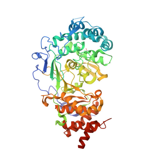
6HMK

POLYADPRIBOSYL GLYCOHYDROLASE IN COMPLEX WITH PDD00016690


Experimental Data Snapshot

  • Method: X-RAY DIFFRACTION
  • Resolution: 2.06 Å
  • R-Value Free: 0.189 
  • R-Value Work: 0.154 
  • R-Value Observed: 0.155 

Starting Model: experimental
View more details

wwPDB Validation   3D Report Full Report


Ligand Structure Quality Assessment 


This is version 1.3 of the entry. See complete history


Literature

Cell-Active Small Molecule Inhibitors of the DNA-Damage Repair Enzyme Poly(ADP-ribose) Glycohydrolase (PARG): Discovery and Optimization of Orally Bioavailable Quinazolinedione Sulfonamides.

Waszkowycz, B.Smith, K.M.McGonagle, A.E.Jordan, A.M.Acton, B.Fairweather, E.E.Griffiths, L.A.Hamilton, N.M.Hamilton, N.S.Hitchin, J.R.Hutton, C.P.James, D.I.Jones, C.D.Jones, S.Mould, D.P.Small, H.F.Stowell, A.I.J.Tucker, J.A.Waddell, I.D.Ogilvie, D.J.

(2018) J Med Chem 61: 10767-10792

  • DOI: https://doi.org/10.1021/acs.jmedchem.8b01407
  • Primary Citation of Related Structures:  
    6HMK, 6HML, 6HMM, 6HMN

  • PubMed Abstract: 

    DNA damage repair enzymes are promising targets in the development of new therapeutic agents for a wide range of cancers and potentially other diseases. The enzyme poly(ADP-ribose) glycohydrolase (PARG) plays a pivotal role in the regulation of DNA repair mechanisms; however, the lack of potent drug-like inhibitors for use in cellular and in vivo models has limited the investigation of its potential as a novel therapeutic target. Using the crystal structure of human PARG in complex with the weakly active and cytotoxic anthraquinone 8a, novel quinazolinedione sulfonamides PARG inhibitors have been identified by means of structure-based virtual screening and library design. 1-Oxetan-3-ylmethyl derivatives 33d and 35d were selected for preliminary investigations in vivo. X-ray crystal structures help rationalize the observed structure-activity relationships of these novel inhibitors.


  • Organizational Affiliation

    Cancer Research UK Manchester Institute , The University of Manchester , Alderley Park , Maccelsfield SK10 4TG , U.K.


Macromolecules
Find similar proteins by:  (by identity cutoff)  |  3D Structure
Entity ID: 1
MoleculeChains Sequence LengthOrganismDetailsImage
Poly(ADP-ribose) glycohydrolase531Homo sapiensMutation(s): 6 
Gene Names: PARG
EC: 3.2.1.143
UniProt & NIH Common Fund Data Resources
Find proteins for Q86W56 (Homo sapiens)
Explore Q86W56 
Go to UniProtKB:  Q86W56
PHAROS:  Q86W56
GTEx:  ENSG00000227345 
Entity Groups  
Sequence Clusters30% Identity50% Identity70% Identity90% Identity95% Identity100% Identity
UniProt GroupQ86W56
Sequence Annotations
Expand
  • Reference Sequence
Small Molecules
Ligands 4 Unique
IDChains Name / Formula / InChI Key2D Diagram3D Interactions
7JC (Subject of Investigation/LOI)
Query on 7JC

Download Ideal Coordinates CCD File 
I [auth A]1-methyl-~{N}-(1-methylcyclopropyl)-3-[(2-methyl-1,3-thiazol-5-yl)methyl]-2,4-bis(oxidanylidene)quinazoline-6-sulfonamide
C18 H20 N4 O4 S2
CZHJSHVWPOJSIU-UHFFFAOYSA-N
SO4
Query on SO4

Download Ideal Coordinates CCD File 
E [auth A],
F [auth A],
G [auth A],
H [auth A]
SULFATE ION
O4 S
QAOWNCQODCNURD-UHFFFAOYSA-L
GOL
Query on GOL

Download Ideal Coordinates CCD File 
D [auth A]GLYCEROL
C3 H8 O3
PEDCQBHIVMGVHV-UHFFFAOYSA-N
DMS
Query on DMS

Download Ideal Coordinates CCD File 
B [auth A],
C [auth A]
DIMETHYL SULFOXIDE
C2 H6 O S
IAZDPXIOMUYVGZ-UHFFFAOYSA-N
Modified Residues  1 Unique
IDChains TypeFormula2D DiagramParent
6WK
Query on 6WK
A
L-PEPTIDE LINKINGC7 H15 N O4 S3CYS
Binding Affinity Annotations 
IDSourceBinding Affinity
7JC BindingDB:  6HMK IC50: 483 (nM) from 1 assay(s)
EC50: 480 (nM) from 1 assay(s)
Experimental Data & Validation

Experimental Data

  • Method: X-RAY DIFFRACTION
  • Resolution: 2.06 Å
  • R-Value Free: 0.189 
  • R-Value Work: 0.154 
  • R-Value Observed: 0.155 
  • Space Group: P 21 21 21
Unit Cell:
Length ( Å )Angle ( ˚ )
a = 67.09α = 90
b = 90.11β = 90
c = 95.5γ = 90
Software Package:
Software NamePurpose
XDSdata reduction
Aimlessdata scaling
BUSTERrefinement
PHASERphasing

Structure Validation

View Full Validation Report


Ligand Structure Quality Assessment 


Entry History 

Deposition Data

Revision History  (Full details and data files)

  • Version 1.0: 2018-11-14
    Type: Initial release
  • Version 1.1: 2018-11-21
    Changes: Data collection, Database references
  • Version 1.2: 2019-04-24
    Changes: Data collection, Database references
  • Version 1.3: 2024-01-24
    Changes: Data collection, Database references, Refinement description