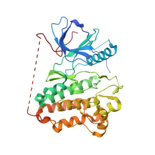
5Y9T

Crystal Structure of EGFR T790M mutant in complex with naquotinib


Experimental Data Snapshot

  • Method: X-RAY DIFFRACTION
  • Resolution: 3.25 Å
  • R-Value Free: 0.240 
  • R-Value Work: 0.195 
  • R-Value Observed: 0.197 

Starting Model: experimental
View more details

wwPDB Validation   3D Report Full Report


Ligand Structure Quality Assessment 


This is version 1.2 of the entry. See complete history


Literature

Pharmacological and Structural Characterizations of Naquotinib, a Novel Third-Generation EGFR Tyrosine Kinase Inhibitor, inEGFR-Mutated Non-Small Cell Lung Cancer.

Hirano, T.Yasuda, H.Hamamoto, J.Nukaga, S.Masuzawa, K.Kawada, I.Naoki, K.Niimi, T.Mimasu, S.Sakagami, H.Soejima, K.Betsuyaku, T.

(2018) Mol Cancer Ther 17: 740-750

  • DOI: https://doi.org/10.1158/1535-7163.MCT-17-1033
  • Primary Citation of Related Structures:  
    5Y9T

  • PubMed Abstract: 

    Multiple epidermal growth factor receptor (EGFR) tyrosine kinase inhibitors (EGFR-TKI) have been developed to effectively inhibit EGFR-derived signals in non-small cell lung cancer (NSCLC). In this study, we assessed the efficacy of EGFR-TKIs, including a novel third-generation inhibitor naquotinib (ASP8273), in clinically relevant EGFR mutations, including L858R, exon 19 deletion, L858R+T790M, exon 19 deletion+T790M with or without a C797S mutation, and several exon 20 insertion mutations. Using structural analyses, we also elucidated the mechanism of activation and sensitivity/resistance to EGFR-TKIs in EGFR exon 20 insertion mutations. The efficacy of naquotinib in cells with L858R, exon 19 deletion and exon 19 deletion+T790M was comparable with that of osimertinib. Interestingly, naquotinib was more potent than osimertinib for L858R+T790M. Additionally, naquotinib and osimertinib had comparable efficacy and a wide therapeutic window for cells with EGFR exon 20 insertions. Structural modeling partly elucidated the mechanism of activation and sensitivity/resistance to EGFR-TKIs in two EGFR exon 20 insertion mutants, A767_V769dupASV and Y764_V765insHH. In summary, we have characterized the efficacy of EGFR-TKIs for NSCLC using in vitro and structural analyses and suggested the mechanism of activation and resistance to EGFR-TKIs of EGFR exon 20 insertion mutations. Our findings should guide the selection of appropriate EGFR-TKIs for the treatment of NSCLC with EGFR mutations and help clarify the biology of EGFR exon 20 insertion mutations. Mol Cancer Ther; 17(4); 740-50. ©2018 AACR .


  • Organizational Affiliation

    Division of Pulmonary Medicine, Department of Medicine, Keio University, School of Medicine, Tokyo, Japan.


Macromolecules
Find similar proteins by:  (by identity cutoff)  |  3D Structure
Entity ID: 1
MoleculeChains Sequence LengthOrganismDetailsImage
Epidermal growth factor receptor333Homo sapiensMutation(s): 1 
Gene Names: EGFRERBBERBB1HER1
EC: 2.7.10.1
UniProt & NIH Common Fund Data Resources
Find proteins for P00533 (Homo sapiens)
Explore P00533 
Go to UniProtKB:  P00533
PHAROS:  P00533
GTEx:  ENSG00000146648 
Entity Groups  
Sequence Clusters30% Identity50% Identity70% Identity90% Identity95% Identity100% Identity
UniProt GroupP00533
Sequence Annotations
Expand
  • Reference Sequence
Small Molecules
Ligands 1 Unique
IDChains Name / Formula / InChI Key2D Diagram3D Interactions
8RC
Query on 8RC

Download Ideal Coordinates CCD File 
B [auth A]6-ethyl-3-[[4-[4-(4-methylpiperazin-1-yl)piperidin-1-yl]phenyl]amino]-5-[(3R)-1-prop-2-enoylpyrrolidin-3-yl]oxy-pyrazin e-2-carboxamide
C30 H42 N8 O3
QKDCLUARMDUUKN-XMMPIXPASA-N
Binding Affinity Annotations 
IDSourceBinding Affinity
8RC BindingDB:  5Y9T IC50: min: 1.7, max: 230 (nM) from 4 assay(s)
Experimental Data & Validation

Experimental Data

  • Method: X-RAY DIFFRACTION
  • Resolution: 3.25 Å
  • R-Value Free: 0.240 
  • R-Value Work: 0.195 
  • R-Value Observed: 0.197 
  • Space Group: I 2 3
Unit Cell:
Length ( Å )Angle ( ˚ )
a = 144.64α = 90
b = 144.64β = 90
c = 144.64γ = 90
Software Package:
Software NamePurpose
REFMACrefinement
HKL-2000data reduction
SCALEPACKdata scaling
PHASERphasing

Structure Validation

View Full Validation Report


Ligand Structure Quality Assessment 


Entry History 

Deposition Data

Revision History  (Full details and data files)

  • Version 1.0: 2018-07-11
    Type: Initial release
  • Version 1.1: 2023-11-22
    Changes: Data collection, Database references, Derived calculations, Refinement description, Structure summary
  • Version 1.2: 2024-10-30
    Changes: Structure summary