5NUR

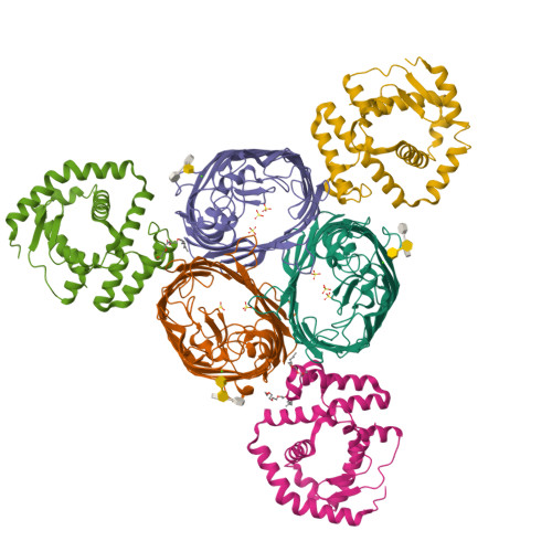
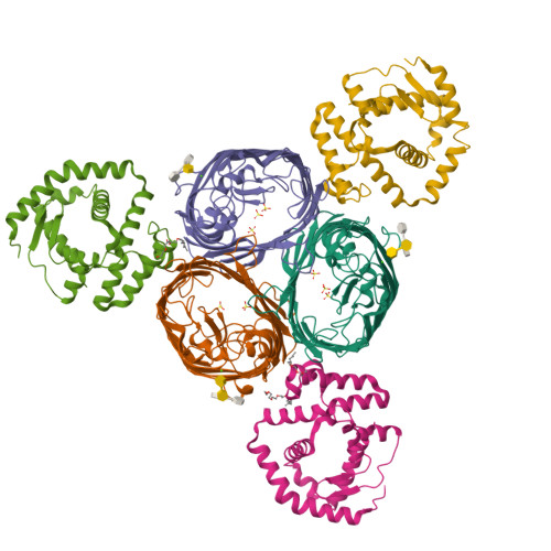
Structural basis for maintenance of bacterial outer membrane lipid asymmetry


Experimental Data Snapshot

  • Method: X-RAY DIFFRACTION
  • Resolution: 3.29 Å
  • R-Value Free: 0.259 
  • R-Value Work: 0.219 
  • R-Value Observed: 0.221 

Starting Model: experimental
View more details

wwPDB Validation   3D Report Full Report


This is version 2.1 of the entry. See complete history


Literature

Structural basis for maintenance of bacterial outer membrane lipid asymmetry.

Abellon-Ruiz, J.Kaptan, S.S.Basle, A.Claudi, B.Bumann, D.Kleinekathofer, U.van den Berg, B.

(2017) Nat Microbiol 2: 1616-1623

  • DOI: https://doi.org/10.1038/s41564-017-0046-x
  • Primary Citation of Related Structures:  
    5NUO, 5NUP, 5NUQ, 5NUR

  • PubMed Abstract: 

    The Gram-negative bacterial outer membrane (OM) is a unique bilayer that forms an efficient permeation barrier to protect the cell from noxious compounds 1 , 2 . The defining characteristic of the OM is lipid asymmetry, with phospholipids comprising the inner leaflet and lipopolysaccharides comprising the outer leaflet 1-3 . This asymmetry is maintained by the Mla pathway, a six-component system that is widespread in Gram-negative bacteria and is thought to mediate retrograde transport of misplaced phospholipids from the outer leaflet of the OM to the cytoplasmic membrane 4 . The OM lipoprotein MlaA performs the first step in this process via an unknown mechanism that does not require external energy input. Here we show, using X-ray crystallography, molecular dynamics simulations and in vitro and in vivo functional assays, that MlaA is a monomeric α-helical OM protein that functions as a phospholipid translocation channel, forming a ~20-Å-thick doughnut embedded in the inner leaflet of the OM with a central, amphipathic pore. This architecture prevents access of inner leaflet phospholipids to the pore, but allows outer leaflet phospholipids to bind to a pronounced ridge surrounding the channel, followed by diffusion towards the periplasmic space. Enterobacterial MlaA proteins form stable complexes with OmpF/C 5,6 , but the porins do not appear to play an active role in phospholipid transport. MlaA represents a lipid transport protein that selectively removes outer leaflet phospholipids to help maintain the essential barrier function of the bacterial OM.


  • Organizational Affiliation

    Institute for Cell and Molecular Biosciences, The Medical School, Newcastle University, Newcastle upon Tyne, NE2 4HH, UK.


Macromolecules
Find similar proteins by:  (by identity cutoff)  |  3D Structure
Entity ID: 1
MoleculeChains Sequence LengthOrganismDetailsImage
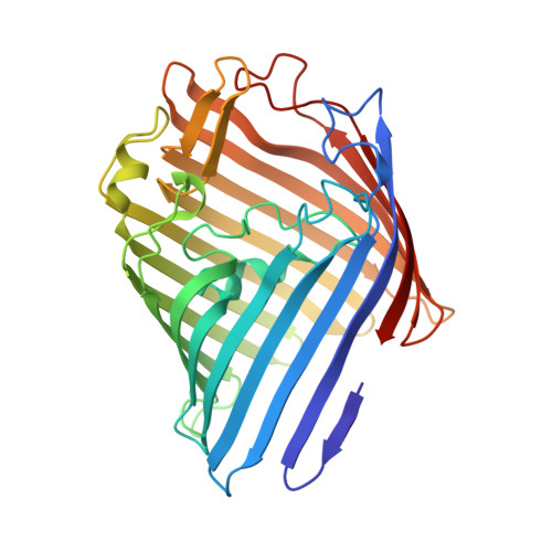
Outer membrane protein FA [auth C],
C [auth A],
E
340Escherichia coli K-12Mutation(s): 0 
Gene Names: ompFcmlBcoacrytolFb0929JW0912
Membrane Entity: Yes 
UniProt
Find proteins for P02931 (Escherichia coli (strain K12))
Explore P02931 
Go to UniProtKB:  P02931
Entity Groups  
Sequence Clusters30% Identity50% Identity70% Identity90% Identity95% Identity100% Identity
UniProt GroupP02931
Sequence Annotations
Expand
  • Reference Sequence
Find similar proteins by:  (by identity cutoff)  |  3D Structure
Entity ID: 2
MoleculeChains Sequence LengthOrganismDetailsImage
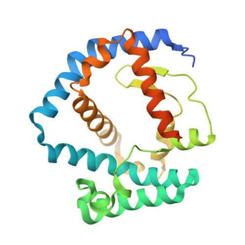
ABC transporter permeaseB [auth D],
D [auth B],
F
236Klebsiella pneumoniaeMutation(s): 0 
Gene Names: 
UniProt
Find proteins for A0A0W8AQT6 (Klebsiella pneumoniae)
Explore A0A0W8AQT6 
Go to UniProtKB:  A0A0W8AQT6
Entity Groups  
Sequence Clusters30% Identity50% Identity70% Identity90% Identity95% Identity100% Identity
UniProt GroupA0A0W8AQT6
Sequence Annotations
Expand
  • Reference Sequence
Oligosaccharides

Help

Entity ID: 3
MoleculeChains Length2D Diagram Glycosylation3D Interactions
3-deoxy-alpha-D-manno-oct-2-ulopyranosonic acid-(2-4)-3-deoxy-alpha-D-manno-oct-2-ulopyranosonic acid-(2-6)-2-amino-2-deoxy-4-O-phosphono-beta-D-glucopyranose-(1-6)-2-amino-2-deoxy-1-O-phosphono-alpha-D-glucopyranose
G
4N/A
Entity ID: 4
MoleculeChains Length2D Diagram Glycosylation3D Interactions
3-deoxy-alpha-D-manno-oct-2-ulopyranosonic acid-(3-4)-3-deoxy-alpha-D-manno-oct-2-ulopyranosonic acid-(2-6)-2-amino-2-deoxy-4-O-phosphono-beta-D-glucopyranose-(1-6)-2-amino-2-deoxy-1-O-phosphono-alpha-D-glucopyranose
H
4N/A
Entity ID: 5
MoleculeChains Length2D Diagram Glycosylation3D Interactions
3-deoxy-alpha-D-manno-oct-2-ulopyranosonic acid-(3-6)-2-amino-2-deoxy-4-O-phosphono-beta-D-glucopyranose-(1-6)-2-amino-2-deoxy-1-O-phosphono-alpha-D-glucopyranose
I
3N/A
Small Molecules
Ligands 3 Unique
IDChains Name / Formula / InChI Key2D Diagram3D Interactions
C8E
Query on C8E

Download Ideal Coordinates CCD File 
N [auth A],
O [auth A],
S [auth E]
(HYDROXYETHYLOXY)TRI(ETHYLOXY)OCTANE
C16 H34 O5
FEOZZFHAVXYAMB-UHFFFAOYSA-N
SO4
Query on SO4

Download Ideal Coordinates CCD File 
K [auth C]
L [auth C]
M [auth C]
Q [auth A]
R [auth A]
K [auth C],
L [auth C],
M [auth C],
Q [auth A],
R [auth A],
U [auth E],
V [auth E],
W [auth E]
SULFATE ION
O4 S
QAOWNCQODCNURD-UHFFFAOYSA-L
CA
Query on CA

Download Ideal Coordinates CCD File 
J [auth C],
P [auth A],
T [auth E]
CALCIUM ION
Ca
BHPQYMZQTOCNFJ-UHFFFAOYSA-N
Experimental Data & Validation

Experimental Data

  • Method: X-RAY DIFFRACTION
  • Resolution: 3.29 Å
  • R-Value Free: 0.259 
  • R-Value Work: 0.219 
  • R-Value Observed: 0.221 
  • Space Group: C 1 2 1
Unit Cell:
Length ( Å )Angle ( ˚ )
a = 190.55α = 90
b = 179.66β = 116.55
c = 133.39γ = 90
Software Package:
Software NamePurpose
XDSdata reduction
xia2data reduction
Aimlessdata scaling
PHASERphasing
PHENIXrefinement
Cootmodel building

Structure Validation

View Full Validation Report


Entry History & Funding Information

Deposition Data


Funding OrganizationLocationGrant Number
European UnionUnited Kingdom115525

Revision History  (Full details and data files)

  • Version 1.0: 2017-10-25
    Type: Initial release
  • Version 1.1: 2017-11-01
    Changes: Database references
  • Version 1.2: 2017-12-06
    Changes: Database references
  • Version 2.0: 2020-07-29
    Type: Remediation
    Reason: Carbohydrate remediation
    Changes: Advisory, Atomic model, Data collection, Derived calculations, Non-polymer description, Structure summary
  • Version 2.1: 2024-01-31
    Changes: Data collection, Database references, Refinement description