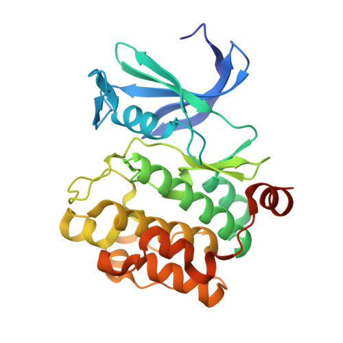
5KCX

Pim-1 kinase in Complex with a Selective N-substituted 7-azaindole Inhibitor


Experimental Data Snapshot

  • Method: X-RAY DIFFRACTION
  • Resolution: 2.20 Å
  • R-Value Free: 0.211 
  • R-Value Work: 0.190 

wwPDB Validation   3D Report Full Report


Ligand Structure Quality Assessment 


This is version 1.2 of the entry. See complete history


Literature

Discovery of N-substituted 7-azaindoles as PIM1 kinase inhibitors - Part I.

Barberis, C.Moorcroft, N.Arendt, C.Levit, M.Moreno-Mazza, S.Batchelor, J.Mechin, I.Majid, T.

(2017) Bioorg Med Chem Lett 27: 4730-4734

  • DOI: https://doi.org/10.1016/j.bmcl.2017.08.069
  • Primary Citation of Related Structures:  
    5KCX, 5TEL, 5TEX, 5TOE, 5TUR

  • PubMed Abstract: 

    Novel N-substituted azaindoles have been discovered as PIM1 inhibitors. X-ray structures have played a significant role in orienting the chemistry effort in the initial phase of hit confirmation. Disclosure of an unconventional binding mode for 1 and 2, as demonstrated by X-ray crystallography, is presented and was an important factor in selecting and advancing a lead series.


  • Organizational Affiliation

    IDD Medicinal Chemistry, Sanofi Genzyme, 153 Second Avenue, Waltham MA 02451, USA. Electronic address: [email protected].


Macromolecules
Find similar proteins by:  (by identity cutoff)  |  3D Structure
Entity ID: 1
MoleculeChains Sequence LengthOrganismDetailsImage
Serine/threonine-protein kinase pim-1291Homo sapiensMutation(s): 0 
Gene Names: PIM1
EC: 2.7.11.1
UniProt & NIH Common Fund Data Resources
Find proteins for P11309 (Homo sapiens)
Explore P11309 
Go to UniProtKB:  P11309
PHAROS:  P11309
GTEx:  ENSG00000137193 
Entity Groups  
Sequence Clusters30% Identity50% Identity70% Identity90% Identity95% Identity100% Identity
UniProt GroupP11309
Sequence Annotations
Expand
  • Reference Sequence
Small Molecules
Binding Affinity Annotations 
IDSourceBinding Affinity
6S3 BindingDB:  5KCX IC50: min: 200, max: 250 (nM) from 2 assay(s)
Experimental Data & Validation

Experimental Data

  • Method: X-RAY DIFFRACTION
  • Resolution: 2.20 Å
  • R-Value Free: 0.211 
  • R-Value Work: 0.190 
  • Space Group: P 65
Unit Cell:
Length ( Å )Angle ( ˚ )
a = 98.01α = 90
b = 98.01β = 90
c = 80.67γ = 120
Software Package:
Software NamePurpose
CNXrefinement
HKL-2000data reduction
HKL-2000data scaling
CNXphasing

Structure Validation

View Full Validation Report


Ligand Structure Quality Assessment 


Entry History 

Deposition Data

Revision History  (Full details and data files)

  • Version 1.0: 2017-07-19
    Type: Initial release
  • Version 1.1: 2017-10-18
    Changes: Database references
  • Version 1.2: 2024-03-06
    Changes: Data collection, Database references