5A3N

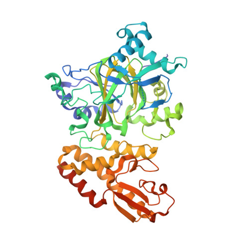
Crystal structure of human PLU-1 (JARID1B) in complex with KDOAM25a


Experimental Data Snapshot

  • Method: X-RAY DIFFRACTION
  • Resolution: 2.00 Å
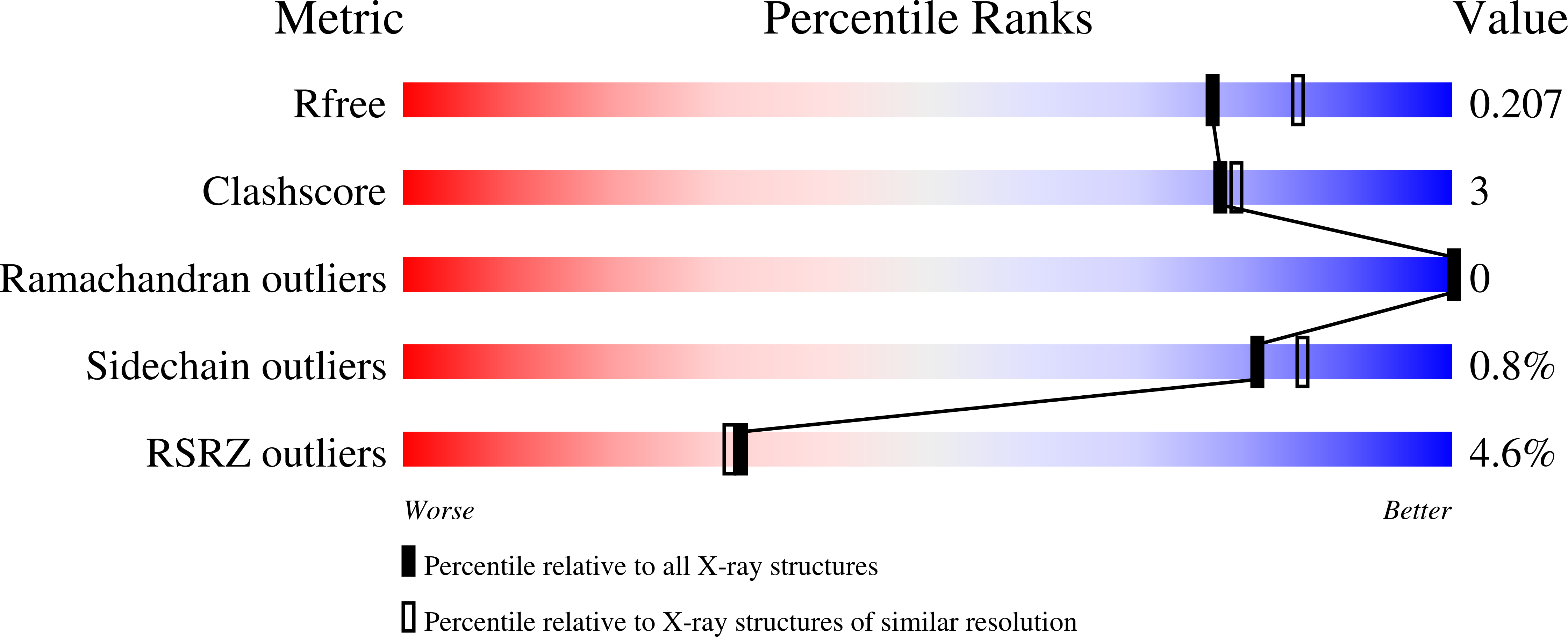
  • R-Value Free: 0.203 
  • R-Value Work: 0.183 
  • R-Value Observed: 0.184 

wwPDB Validation   3D Report Full Report


Ligand Structure Quality Assessment 


This is version 2.0 of the entry. See complete history


Literature

Potent and Selective KDM5 Inhibitor Stops Cellular Demethylation of H3K4me3 at Transcription Start Sites and Proliferation of MM1S Myeloma Cells.

Tumber, A.Nuzzi, A.Hookway, E.S.Hatch, S.B.Velupillai, S.Johansson, C.Kawamura, A.Savitsky, P.Yapp, C.Szykowska, A.Wu, N.Bountra, C.Strain-Damerell, C.Burgess-Brown, N.A.Ruda, G.F.Fedorov, O.Munro, S.England, K.S.Nowak, R.P.Schofield, C.J.La Thangue, N.B.Pawlyn, C.Davies, F.Morgan, G.Athanasou, N.Muller, S.Oppermann, U.Brennan, P.E.

(2017) Cell Chem Biol 24: 371-380

  • DOI: https://doi.org/10.1016/j.chembiol.2017.02.006
  • Primary Citation of Related Structures:  
    5A3N

  • PubMed Abstract: 

    Methylation of lysine residues on histone tail is a dynamic epigenetic modification that plays a key role in chromatin structure and gene regulation. Members of the KDM5 (also known as JARID1) sub-family are 2-oxoglutarate (2-OG) and Fe 2+ -dependent oxygenases acting as histone 3 lysine 4 trimethyl (H3K4me3) demethylases, regulating proliferation, stem cell self-renewal, and differentiation. Here we present the characterization of KDOAM-25, an inhibitor of KDM5 enzymes. KDOAM-25 shows biochemical half maximal inhibitory concentration values of <100 nM for KDM5A-D in vitro, high selectivity toward other 2-OG oxygenases sub-families, and no off-target activity on a panel of 55 receptors and enzymes. In human cell assay systems, KDOAM-25 has a half maximal effective concentration of ∼50 μM and good selectivity toward other demethylases. KDM5B is overexpressed in multiple myeloma and negatively correlated with the overall survival. Multiple myeloma MM1S cells treated with KDOAM-25 show increased global H3K4 methylation at transcriptional start sites and impaired proliferation.


  • Organizational Affiliation

    Structural Genomics Consortium, University of Oxford, Oxford OX3 7DQ, UK; Nuffield Department of Medicine, Target Discovery Institute, University of Oxford, Oxford OX3 7FZ, UK.


Macromolecules
Find similar proteins by:  (by identity cutoff)  |  3D Structure
Entity ID: 1
MoleculeChains Sequence LengthOrganismDetailsImage
LYSINE-SPECIFIC DEMETHYLASE 5B481Homo sapiensMutation(s): 0 
EC: 1.14.11 (PDB Primary Data), 1.14.11.67 (UniProt)
UniProt & NIH Common Fund Data Resources
Find proteins for Q9UGL1 (Homo sapiens)
Explore Q9UGL1 
Go to UniProtKB:  Q9UGL1
PHAROS:  Q9UGL1
GTEx:  ENSG00000117139 
Entity Groups  
Sequence Clusters30% Identity50% Identity70% Identity90% Identity95% Identity100% Identity
UniProt GroupQ9UGL1
Sequence Annotations
Expand
  • Reference Sequence
Small Molecules
Ligands 7 Unique
IDChains Name / Formula / InChI Key2D Diagram3D Interactions
LQT
Query on LQT

Download Ideal Coordinates CCD File 
G [auth A]2-[[[2-[2-(dimethylamino)ethyl-ethyl-amino]-2-oxidanylidene-ethyl]amino]methyl]pyridine-4-carboxamide
C15 H25 N5 O2
PNFMVADNCOGWME-UHFFFAOYSA-N
EPE
Query on EPE

Download Ideal Coordinates CCD File 
J [auth A]4-(2-HYDROXYETHYL)-1-PIPERAZINE ETHANESULFONIC ACID
C8 H18 N2 O4 S
JKMHFZQWWAIEOD-UHFFFAOYSA-N
PO4
Query on PO4

Download Ideal Coordinates CCD File 
I [auth A]PHOSPHATE ION
O4 P
NBIIXXVUZAFLBC-UHFFFAOYSA-K
DMS
Query on DMS

Download Ideal Coordinates CCD File 
H [auth A]DIMETHYL SULFOXIDE
C2 H6 O S
IAZDPXIOMUYVGZ-UHFFFAOYSA-N
ZN
Query on ZN

Download Ideal Coordinates CCD File 
B [auth A],
C [auth A]
ZINC ION
Zn
PTFCDOFLOPIGGS-UHFFFAOYSA-N
EDO
Query on EDO

Download Ideal Coordinates CCD File 
K [auth A]
L [auth A]
M [auth A]
N [auth A]
O [auth A]
K [auth A],
L [auth A],
M [auth A],
N [auth A],
O [auth A],
P [auth A]
1,2-ETHANEDIOL
C2 H6 O2
LYCAIKOWRPUZTN-UHFFFAOYSA-N
MN
Query on MN

Download Ideal Coordinates CCD File 
D [auth A],
E [auth A],
F [auth A]
MANGANESE (II) ION
Mn
WAEMQWOKJMHJLA-UHFFFAOYSA-N
Binding Affinity Annotations 
IDSourceBinding Affinity
LQT BindingDB:  5A3N IC50: min: 19, max: 558 (nM) from 4 assay(s)
Experimental Data & Validation

Experimental Data

  • Method: X-RAY DIFFRACTION
  • Resolution: 2.00 Å
  • R-Value Free: 0.203 
  • R-Value Work: 0.183 
  • R-Value Observed: 0.184 
  • Space Group: P 65 2 2
Unit Cell:
Length ( Å )Angle ( ˚ )
a = 141.874α = 90
b = 141.874β = 90
c = 151.865γ = 120
Software Package:
Software NamePurpose
REFMACrefinement
XDSdata reduction
SCALAdata scaling
CCP4-AUTOMRphasing

Structure Validation

View Full Validation Report


Ligand Structure Quality Assessment 


Entry History 

Revision History  (Full details and data files)

  • Version 1.0: 2015-07-08
    Type: Initial release
  • Version 1.1: 2017-03-22
    Changes: Database references
  • Version 1.2: 2017-03-29
    Changes: Database references
  • Version 2.0: 2024-05-08
    Changes: Atomic model, Data collection, Database references, Derived calculations, Other