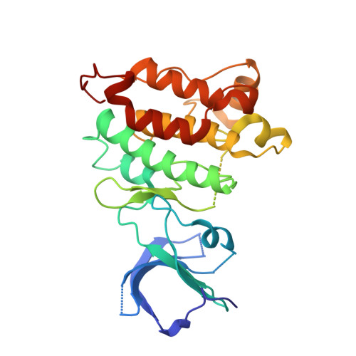
4LGH

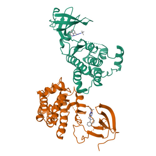
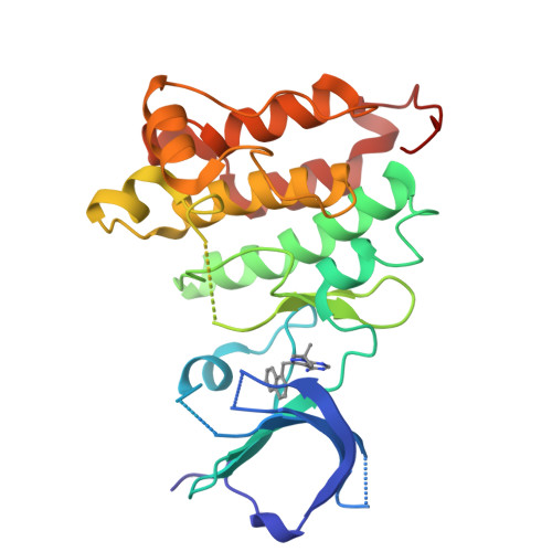
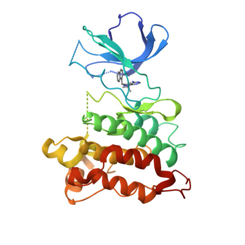
Crystal structure of 1NM-PP1 bound to analog-sensitive Src kinase


Experimental Data Snapshot

  • Method: X-RAY DIFFRACTION
  • Resolution: 2.84 Å
  • R-Value Free: 0.272 
  • R-Value Work: 0.213 
  • R-Value Observed: 0.216 

Starting Model: experimental
View more details

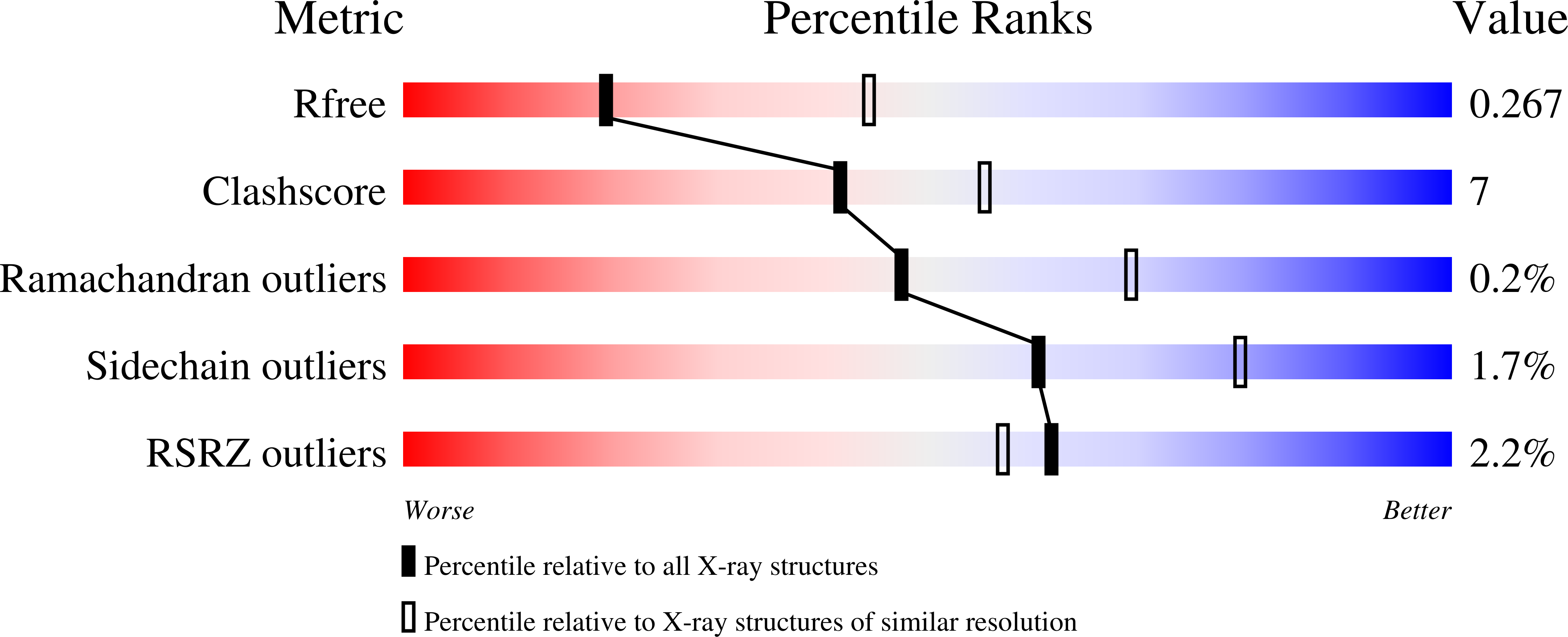
wwPDB Validation   3D Report Full Report


Ligand Structure Quality Assessment 


This is version 1.3 of the entry. See complete history


Literature

Structure-guided inhibitor design expands the scope of analog-sensitive kinase technology.

Zhang, C.Lopez, M.S.Dar, A.C.Ladow, E.Finkbeiner, S.Yun, C.H.Eck, M.J.Shokat, K.M.

(2013) ACS Chem Biol 8: 1931-1938

  • DOI: https://doi.org/10.1021/cb400376p
  • Primary Citation of Related Structures:  
    4LGG, 4LGH

  • PubMed Abstract: 

    Engineered analog-sensitive (AS) protein kinases have emerged as powerful tools for dissecting phospho-signaling pathways, for elucidating the cellular function of individual kinases, and for deciphering unanticipated effects of clinical therapeutics. A crucial and necessary feature of this technology is a bioorthogonal small molecule that is innocuous toward native cellular systems but potently inhibits the engineered kinase. In order to generalize this method, we sought a molecule capable of targeting divergent AS-kinases. Here we employ X-ray crystallography and medicinal chemistry to unravel the mechanism of current inhibitors and use these insights to design the most potent, selective, and general AS-kinase inhibitors reported to date. We use large-scale kinase inhibitor profiling to characterize the selectivity of these molecules as well as examine the consequences of potential off-target effects in chemical genetic experiments. The molecules reported here will serve as powerful tools in efforts to extend AS-kinase technology to the entire kinome and the principles discovered may help in the design of other engineered enzyme/ligand pairs.


  • Organizational Affiliation

    Howard Hughes Medical Institute and Department of Cellular & Molecular Pharmacology, University of California, San Francisco , San Francisco, California 94158, United States.


Macromolecules
Find similar proteins by:  (by identity cutoff)  |  3D Structure
Entity ID: 1
MoleculeChains Sequence LengthOrganismDetailsImage
Proto-oncogene tyrosine-protein kinase Src
A, B
277Gallus gallusMutation(s): 1 
Gene Names: SRC
EC: 2.7.10.2
UniProt
Find proteins for P00523 (Gallus gallus)
Explore P00523 
Go to UniProtKB:  P00523
Entity Groups  
Sequence Clusters30% Identity50% Identity70% Identity90% Identity95% Identity100% Identity
UniProt GroupP00523
Sequence Annotations
Expand
  • Reference Sequence
Small Molecules
Ligands 1 Unique
IDChains Name / Formula / InChI Key2D Diagram3D Interactions
0JN
Query on 0JN

Download Ideal Coordinates CCD File 
C [auth A],
D [auth B]
1-tert-butyl-3-(naphthalen-1-ylmethyl)-1H-pyrazolo[3,4-d]pyrimidin-4-amine
C20 H21 N5
GDQXJQSQYMMKRA-UHFFFAOYSA-N
Binding Affinity Annotations 
IDSourceBinding Affinity
0JN BindingDB:  4LGH IC50: min: 620, max: 2200 (nM) from 2 assay(s)
PDBBind:  4LGH IC50: 2 (nM) from 1 assay(s)
Experimental Data & Validation

Experimental Data

  • Method: X-RAY DIFFRACTION
  • Resolution: 2.84 Å
  • R-Value Free: 0.272 
  • R-Value Work: 0.213 
  • R-Value Observed: 0.216 
  • Space Group: P 1
Unit Cell:
Length ( Å )Angle ( ˚ )
a = 42.264α = 100.38
b = 62.654β = 91.01
c = 74.325γ = 90.07
Software Package:
Software NamePurpose
DENZOdata reduction
SCALEPACKdata scaling
PHASERphasing
PHENIXrefinement
PDB_EXTRACTdata extraction
HKL-2000data collection

Structure Validation

View Full Validation Report


Ligand Structure Quality Assessment 


Entry History 

Deposition Data

Revision History  (Full details and data files)

  • Version 1.0: 2013-08-07
    Type: Initial release
  • Version 1.1: 2013-10-09
    Changes: Database references
  • Version 1.2: 2017-11-15
    Changes: Refinement description
  • Version 1.3: 2023-09-20
    Changes: Data collection, Database references, Derived calculations, Refinement description