RCSB PDB - 3UEX: Bovine beta-lactoglobulin complex with stearic acid

 3UEX

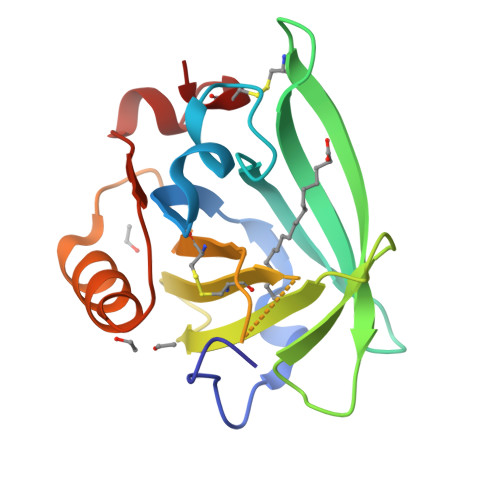
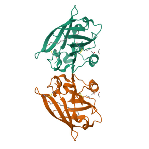
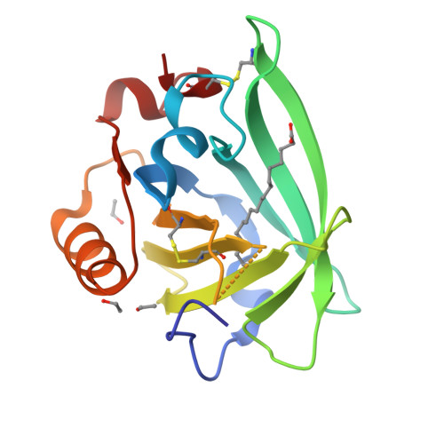
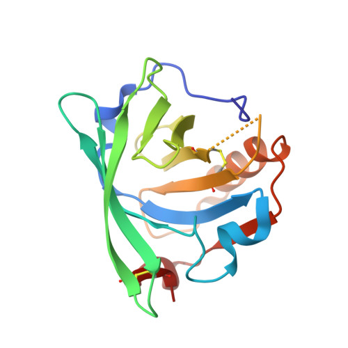
Bovine beta-lactoglobulin complex with stearic acid


Experimental Data Snapshot

  • Method: X-RAY DIFFRACTION
  • Resolution: 2.10 Å
  • R-Value Free: 0.284 
  • R-Value Work: 0.230 
  • R-Value Observed: 0.235 

Starting Model: experimental
View more details

wwPDB Validation   3D Report Full Report


Ligand Structure Quality Assessment 

Created with Raphaël 2.3.0Worse 01 BetterLigand structure goodness of fit to experimental dataBest fitted STEClick on this verticalbar to view details

This is version 1.5 of the entry. See complete history


Literature

Bovine beta-lactoglobulin complex with stearic acid

Loch, J.I.Polit, A.Bonarek, P.Olszewska, D.Kurpiewska, K.Dziedzicka-Wasylewska, M.Lewinski, K.

(2012) Int J Biol Macromol 50: 1095-1102

  • DOI: https://doi.org/10.1016/j.ijbiomac.2012.03.002
  • Primary Citation of Related Structures:  
    3UEU, 3UEV, 3UEW, 3UEX

  • PubMed Abstract: 

    Lactoglobulin is a globular milk protein for which physiological function has not been clarified. Due to its binding properties lactoglobulin might serve as a carrier for bioactive molecules. Binding of 12-, 14-, 16- and 18-carbon saturated fatty acids to bovine β-lactoglobulin has been characterised by isothermal titration calorimetry and X-ray crystallography as a part of systematic studies of lactoglobulin complexes with ligands of biological importance. The thermodynamic parameters have been determined for lauric, myristic and palmitic acid complexes revealing systematic decrease of enthalpic and increase of entropic component of ΔG with elongation of aliphatic chain. In all crystal structures determined with resolution 1.9-2.1Å, single fatty acid molecule was found in the β-barrel in extended conformation with individual pattern of interactions. Location of a fatty acid in the binding site depends on the length of aliphatic chain and influences polar interactions between protein and ligand. Systematic changes of entropic component indicate important role of water in binding process.


  • Organizational Affiliation

    Jagiellonian University, Faculty of Chemistry, Department of Crystal Physics and Crystal Chemistry, Kraków, Poland.


Macromolecules
Find similar proteins by:  (by identity cutoff)  |  3D Structure
Entity ID: 1
MoleculeChains Sequence LengthOrganismDetailsImage
Beta-lactoglobulin162Bos taurusMutation(s): 0 
UniProt
Find proteins for P02754 (Bos taurus)
Explore P02754 
Go to UniProtKB:  P02754
Entity Groups  
Sequence Clusters30% Identity50% Identity70% Identity90% Identity95% Identity100% Identity
UniProt GroupP02754
Sequence Annotations
Expand
  • Reference Sequence
Experimental Data & Validation

Experimental Data

  • Method: X-RAY DIFFRACTION
  • Resolution: 2.10 Å
  • R-Value Free: 0.284 
  • R-Value Work: 0.230 
  • R-Value Observed: 0.235 
  • Space Group: P 32 2 1
Unit Cell:
Length ( Å )Angle ( ˚ )
a = 53.778α = 90
b = 53.778β = 90
c = 112.038γ = 120
Software Package:
Software NamePurpose
CrysalisProdata collection
MOLREPphasing
REFMACrefinement
CrysalisProdata reduction
SCALAdata scaling

Structure Validation

View Full Validation Report


Ligand Structure Quality Assessment 

Created with Raphaël 2.3.0Worse 01 BetterLigand structure goodness of fit to experimental dataBest fitted STEClick on this verticalbar to view details

Entry History 

Deposition Data

Revision History  (Full details and data files)

  • Version 1.0: 2011-11-09
    Type: Initial release
  • Version 1.1: 2012-04-18
    Changes: Database references
  • Version 1.2: 2012-06-20
    Changes: Database references
  • Version 1.3: 2017-11-08
    Changes: Refinement description
  • Version 1.4: 2023-09-13
    Changes: Data collection, Database references, Derived calculations, Refinement description
  • Version 1.5: 2024-10-30
    Changes: Structure summary