3E5A

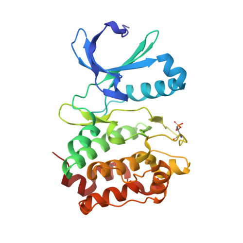
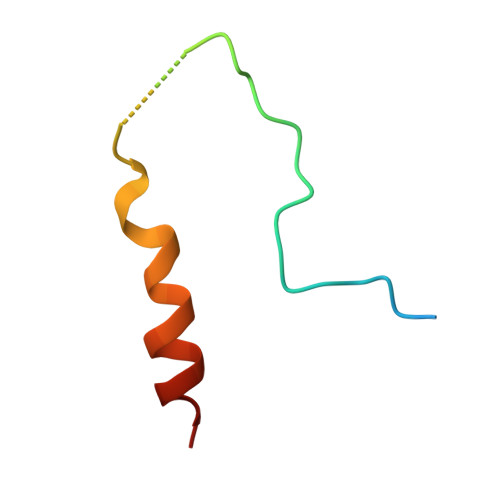
Crystal structure of Aurora A in complex with VX-680 and TPX2


Experimental Data Snapshot

  • Method: X-RAY DIFFRACTION
  • Resolution: 2.30 Å
  • R-Value Free: 0.256 
  • R-Value Work: 0.203 
  • R-Value Observed: 0.206 

Starting Model: experimental
View more details

wwPDB Validation   3D Report Full Report


Ligand Structure Quality Assessment 


This is version 1.3 of the entry. See complete history


Literature

Modulation of kinase-inhibitor interactions by auxiliary protein binding: crystallography studies on Aurora A interactions with VX-680 and with TPX2.

Zhao, B.Smallwood, A.Yang, J.Koretke, K.Nurse, K.Calamari, A.Kirkpatrick, R.B.Lai, Z.

(2008) Protein Sci 17: 1791-1797

  • DOI: https://doi.org/10.1110/ps.036590.108
  • Primary Citation of Related Structures:  
    3E5A

  • PubMed Abstract: 

    VX-680, also known as MK-0457, is an ATP-competitive small molecule inhibitor of the Aurora kinases that has entered phase II clinical trials for the treatment of cancer. We have solved the cocrystal structure of AurA/TPX2/VX-680 at 2.3 A resolution. In the crystal structure, VX-680 binds to the active conformation of AurA. The glycine-rich loop in AurA adopts a unique bent conformation, forming a pi-pi interaction with the phenyl group of VX-680. In contrast, in the published AurA/VX-680 structure, VX-680 binds to AurA in the inactive conformation, interacting with a hydrophobic pocket only present in the inactive conformation. These data suggest that TPX2, a protein cofactor, can alter the binding mode of VX-680 with AurA. More generally, the presence of physiologically relevant cofactor proteins can alter the kinetics, binding interactions, and inhibition of enzymes, and studies with these multiprotein complexes may be beneficial to the discovery and optimization of enzyme inhibitors as therapeutic agents.


  • Organizational Affiliation

    Department of Computational and Structural Chemistry, GlaxoSmithKline, 709 Swedeland Road, King of Prussia, PA 19406, USA. [email protected]


Macromolecules
Find similar proteins by:  (by identity cutoff)  |  3D Structure
Entity ID: 1
MoleculeChains Sequence LengthOrganismDetailsImage
Serine/threonine-protein kinase 6268Homo sapiensMutation(s): 0 
Gene Names: AURKAAIKARK1AURABTAKSTK15STK6
EC: 2.7.11.1
UniProt & NIH Common Fund Data Resources
Find proteins for O14965 (Homo sapiens)
Explore O14965 
Go to UniProtKB:  O14965
PHAROS:  O14965
GTEx:  ENSG00000087586 
Entity Groups  
Sequence Clusters30% Identity50% Identity70% Identity90% Identity95% Identity100% Identity
UniProt GroupO14965
Sequence Annotations
Expand
  • Reference Sequence
Find similar proteins by:  (by identity cutoff)  |  3D Structure
Entity ID: 2
MoleculeChains Sequence LengthOrganismDetailsImage
Targeting protein for Xklp244Homo sapiensMutation(s): 0 
Gene Names: TPX2C20orf1C20orf2DIL2HCA519
UniProt & NIH Common Fund Data Resources
Find proteins for Q9ULW0 (Homo sapiens)
Explore Q9ULW0 
Go to UniProtKB:  Q9ULW0
PHAROS:  Q9ULW0
GTEx:  ENSG00000088325 
Entity Groups  
Sequence Clusters30% Identity50% Identity70% Identity90% Identity95% Identity100% Identity
UniProt GroupQ9ULW0
Sequence Annotations
Expand
  • Reference Sequence
Small Molecules
Ligands 2 Unique
IDChains Name / Formula / InChI Key2D Diagram3D Interactions
VX6
Query on VX6

Download Ideal Coordinates CCD File 
C [auth A]CYCLOPROPANECARBOXYLIC ACID {4-[4-(4-METHYL-PIPERAZIN-1-YL)-6-(5-METHYL-2H-PYRAZOL-3-YLAMINO)-PYRIMIDIN-2-YLSULFANYL]-PHENYL}-AMIDE
C23 H28 N8 O S
GCIKSSRWRFVXBI-UHFFFAOYSA-N
SO4
Query on SO4

Download Ideal Coordinates CCD File 
D [auth A]SULFATE ION
O4 S
QAOWNCQODCNURD-UHFFFAOYSA-L
Modified Residues  1 Unique
IDChains TypeFormula2D DiagramParent
TPO
Query on TPO
A
L-PEPTIDE LINKINGC4 H10 N O6 PTHR
Binding Affinity Annotations 
IDSourceBinding Affinity
VX6 PDBBind:  3E5A Ki: 5.9 (nM) from 1 assay(s)
BindingDB:  3E5A Ki: min: 0.6, max: 2 (nM) from 2 assay(s)
Kd: min: 3.9, max: 17 (nM) from 4 assay(s)
IC50: min: 0.6, max: 4954 (nM) from 19 assay(s)
Experimental Data & Validation

Experimental Data

  • Method: X-RAY DIFFRACTION
  • Resolution: 2.30 Å
  • R-Value Free: 0.256 
  • R-Value Work: 0.203 
  • R-Value Observed: 0.206 
  • Space Group: C 2 2 21
Unit Cell:
Length ( Å )Angle ( ˚ )
a = 87.186α = 90
b = 89.097β = 90
c = 88.499γ = 90
Software Package:
Software NamePurpose
REFMACrefinement
ADSCdata collection
HKL-2000data reduction
SCALEPACKdata scaling
PHASERphasing

Structure Validation

View Full Validation Report


Ligand Structure Quality Assessment 


Entry History 

Deposition Data

Revision History  (Full details and data files)

  • Version 1.0: 2008-10-28
    Type: Initial release
  • Version 1.1: 2011-07-13
    Changes: Advisory, Version format compliance
  • Version 1.2: 2023-08-30
    Changes: Data collection, Database references, Derived calculations, Refinement description
  • Version 1.3: 2024-10-30
    Changes: Structure summary