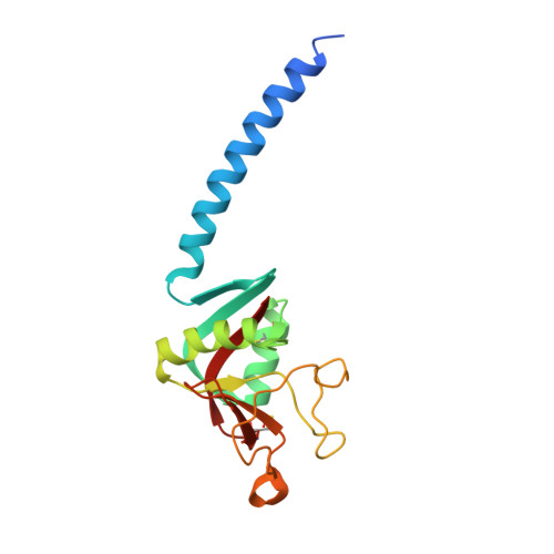
3DBZ

human surfactant protein D


Experimental Data Snapshot

  • Method: X-RAY DIFFRACTION
  • Resolution: 1.80 Å
  • R-Value Free: 0.202 
  • R-Value Work: 0.180 
  • R-Value Observed: 0.182 

Starting Model: experimental
View more details

wwPDB Validation   3D Report Full Report


This is version 1.4 of the entry. See complete history


Literature

Interaction of recombinant surfactant protein D with lipopolysaccharide: conformation and orientation of bound protein by IRRAS and simulations.

Wang, L.Brauner, J.W.Mao, G.Crouch, E.Seaton, B.Head, J.Smith, K.Flach, C.R.Mendelsohn, R.

(2008) Biochemistry 47: 8103-8113

  • DOI: https://doi.org/10.1021/bi800626h
  • Primary Citation of Related Structures:  
    3DBZ

  • PubMed Abstract: 

    Effective innate host defense requires early recognition of pathogens. Surfactant protein D (SP-D), shown to play a role in host defense, binds to the lipopolysaccharide (LPS) component of Gram-negative bacterial membranes. Binding takes place via the carbohydrate recognition domain (CRD) of SP-D. Recombinant trimeric neck+CRDs (NCRD) have proven valuable in biophysical studies of specific interactions. Although X-ray crystallography has provided atomic level information on NCRD binding to carbohydrates and other ligands, molecular level information about interactions between SP-D and biological ligands under physiologically relevant conditions is lacking. Infrared reflection-absorption spectroscopy (IRRAS) provides molecular structure information from films at the air/water interface where protein adsorption to LPS monolayers serves as a model for protein-lipid interaction. In the current studies, we examine the adsorption of NCRDs to Rd 1 LPS monolayers using surface pressure measurements and IRRAS. Measurements of surface pressure, Amide I band intensities, and LPS acyl chain conformational ordering, along with the introduction of EDTA, permit discrimination of Ca (2+)-mediated binding from nonspecific protein adsorption. The findings support the concept of specific binding between the CRD and heptoses in the core region of LPS. In addition, a novel simulation method that accurately predicts the IR Amide I contour from X-ray coordinates of NCRD SP-D is applied and coupled to quantitative IRRAS equations providing information on protein orientation. Marked differences in orientation are found when the NCRD binds to LPS compared to nonspecific adsorption. The geometry suggests that all three CRDs are simultaneously bound to LPS under conditions that support the Ca (2+)-mediated interaction.


  • Organizational Affiliation

    Department of Chemistry, Newark College of Arts and Science, Rutgers University, Newark, New Jersey 07102, USA.


Macromolecules
Find similar proteins by:  (by identity cutoff)  |  3D Structure
Entity ID: 1
MoleculeChains Sequence LengthOrganismDetailsImage
Pulmonary surfactant-associated protein D
A, B, C
160Homo sapiensMutation(s): 0 
Gene Names: SFTPDPSPDSFTP4
UniProt & NIH Common Fund Data Resources
Find proteins for P35247 (Homo sapiens)
Explore P35247 
Go to UniProtKB:  P35247
PHAROS:  P35247
GTEx:  ENSG00000133661 
Entity Groups  
Sequence Clusters30% Identity50% Identity70% Identity90% Identity95% Identity100% Identity
UniProt GroupP35247
Sequence Annotations
Expand
  • Reference Sequence
Small Molecules
Ligands 2 Unique
IDChains Name / Formula / InChI Key2D Diagram3D Interactions
MPD
Query on MPD

Download Ideal Coordinates CCD File 
G [auth A],
H [auth A]
(4S)-2-METHYL-2,4-PENTANEDIOL
C6 H14 O2
SVTBMSDMJJWYQN-YFKPBYRVSA-N
CA
Query on CA

Download Ideal Coordinates CCD File 
D [auth A]
E [auth A]
F [auth A]
I [auth B]
J [auth B]
D [auth A],
E [auth A],
F [auth A],
I [auth B],
J [auth B],
K [auth B],
L [auth C],
M [auth C],
N [auth C]
CALCIUM ION
Ca
BHPQYMZQTOCNFJ-UHFFFAOYSA-N
Experimental Data & Validation

Experimental Data

  • Method: X-RAY DIFFRACTION
  • Resolution: 1.80 Å
  • R-Value Free: 0.202 
  • R-Value Work: 0.180 
  • R-Value Observed: 0.182 
  • Space Group: P 1 21 1
Unit Cell:
Length ( Å )Angle ( ˚ )
a = 55.26α = 90
b = 107.6β = 91.8
c = 55.6γ = 90
Software Package:
Software NamePurpose
DENZOdata reduction
SCALEPACKdata scaling
CNSrefinement
PDB_EXTRACTdata extraction
ADSCdata collection
HKL-2000data reduction
HKL-2000data scaling
CNSphasing

Structure Validation

View Full Validation Report


Entry History 

Deposition Data

  • Released Date: 2008-08-05 
  • Deposition Author(s): Head, J.F.

Revision History  (Full details and data files)

  • Version 1.0: 2008-08-05
    Type: Initial release
  • Version 1.1: 2011-07-13
    Changes: Version format compliance
  • Version 1.2: 2017-10-25
    Changes: Refinement description
  • Version 1.3: 2023-08-30
    Changes: Data collection, Database references, Derived calculations, Refinement description
  • Version 1.4: 2024-11-20
    Changes: Structure summary