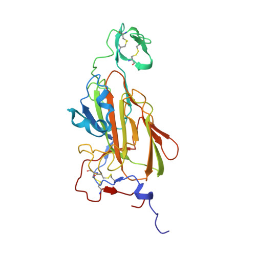
RCSB PDB - 2XJV: X-ray structure of the N-terminal domain of the flocculin Flo5 from Saccharomyces cerevisiae with mutation D201T in complex with calcium and glucose

 2XJV

X-ray structure of the N-terminal domain of the flocculin Flo5 from Saccharomyces cerevisiae with mutation D201T in complex with calcium and glucose


Experimental Data Snapshot

  • Method: X-RAY DIFFRACTION
  • Resolution: 1.74 Å
  • R-Value Free: 0.219 
  • R-Value Work: 0.181 
  • R-Value Observed: 0.182 

Starting Model: other
View more details

wwPDB Validation   3D Report Full Report


Ligand Structure Quality Assessment 

Created with Raphaël 2.3.0Worse 01 BetterLigand structure goodness of fit to experimental dataBest fitted BGCClick on this verticalbar to view details

This is version 1.6 of the entry. See complete history


Literature

Structural Basis of Flocculin-Mediated Social Behavior in Yeast

Veelders, M.Brueckner, S.Ott, D.Unverzagt, C.Moesch, H.-U.Essen, L.-O.

(2010) Proc Natl Acad Sci U S A 107: 22511

  • DOI: https://doi.org/10.1073/pnas.1013210108
  • Primary Citation of Related Structures:  
    2XJP, 2XJQ, 2XJR, 2XJS, 2XJT, 2XJU, 2XJV

  • PubMed Abstract: 

    In the budding yeast Saccharomyces cerevisiae, self-recognition and the thereby promoted aggregation of thousands of cells into protective flocs is mediated by a family of cell-surface adhesins, the flocculins (Flo). Based on this social behavior FLO genes fulfill the definition of "greenbeard" genes, which direct cooperation toward other carriers of the same gene. The process of flocculation plays an eminent role in the food industry for the production of beer and wine. However, the precise mode of flocculin-mediated surface recognition and the exact structure of cognate ligands have remained elusive. Here, we present structures of the adhesion domain of a flocculin complexed to its cognate ligands derived from yeast high-mannose oligosaccharides at resolutions up to 0.95 Å. Besides a PA14-like architecture, the Flo5A domain reveals a previously undescribed lectin fold that utilizes a unique DcisD calcium-binding motif for carbohydrate binding and that is widely spread among pro- and eukaryotes. Given the high abundance of high-mannose oligosaccharides in yeast cell walls, the Flo5A structure suggests a model for recognition, where social non-self- instead of unsocial self-interactions are favored.


  • Organizational Affiliation

    Department of Chemistry/Biochemistry, Philipps-Universität Marburg, Hans-Meerwein-Strasse, D-35032 Marburg, Germany.


Macromolecules
Find similar proteins by:  (by identity cutoff)  |  3D Structure
Entity ID: 1
MoleculeChains Sequence LengthOrganismDetailsImage
FLOCCULATION PROTEIN FLO5258Saccharomyces cerevisiae S288CMutation(s): 1 
UniProt
Find proteins for P38894 (Saccharomyces cerevisiae (strain ATCC 204508 / S288c))
Explore P38894 
Go to UniProtKB:  P38894
Entity Groups  
Sequence Clusters30% Identity50% Identity70% Identity90% Identity95% Identity100% Identity
UniProt GroupP38894
Sequence Annotations
Expand
  • Reference Sequence
Small Molecules
Ligands 4 Unique
IDChains Name / Formula / InChI Key2D Diagram3D Interactions
BGC
Query on BGC

Download Ideal Coordinates CCD File 
G [auth A],
H [auth A]
beta-D-glucopyranose
C6 H12 O6
WQZGKKKJIJFFOK-VFUOTHLCSA-N
CA
Query on CA

Download Ideal Coordinates CCD File 
B [auth A],
C [auth A]
CALCIUM ION
Ca
BHPQYMZQTOCNFJ-UHFFFAOYSA-N
CL
Query on CL

Download Ideal Coordinates CCD File 
E [auth A]CHLORIDE ION
Cl
VEXZGXHMUGYJMC-UHFFFAOYSA-M
NA
Query on NA

Download Ideal Coordinates CCD File 
D [auth A],
F [auth A]
SODIUM ION
Na
FKNQFGJONOIPTF-UHFFFAOYSA-N
Experimental Data & Validation

Experimental Data

  • Method: X-RAY DIFFRACTION
  • Resolution: 1.74 Å
  • R-Value Free: 0.219 
  • R-Value Work: 0.181 
  • R-Value Observed: 0.182 
  • Space Group: P 21 21 21
Unit Cell:
Length ( Å )Angle ( ˚ )
a = 46.45α = 90
b = 62.36β = 90
c = 106.19γ = 90
Software Package:
Software NamePurpose
REFMACrefinement
XDSdata reduction
XSCALEdata scaling
REFMACphasing

Structure Validation

View Full Validation Report


Ligand Structure Quality Assessment 

Created with Raphaël 2.3.0Worse 01 BetterLigand structure goodness of fit to experimental dataBest fitted BGCClick on this verticalbar to view details

Entry History 

Deposition Data

Revision History  (Full details and data files)

  • Version 1.0: 2010-12-15
    Type: Initial release
  • Version 1.1: 2011-05-08
    Changes: Version format compliance
  • Version 1.2: 2011-07-13
    Changes: Version format compliance
  • Version 1.3: 2017-07-05
    Changes: Data collection
  • Version 1.4: 2020-07-29
    Type: Remediation
    Reason: Carbohydrate remediation
    Changes: Data collection, Derived calculations, Other, Structure summary
  • Version 1.5: 2024-05-01
    Changes: Data collection, Database references, Refinement description, Structure summary
  • Version 1.6: 2024-11-06
    Changes: Structure summary