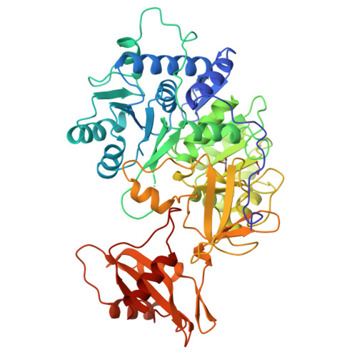
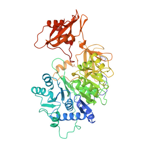
1BA3

FIREFLY LUCIFERASE IN COMPLEX WITH BROMOFORM


Experimental Data Snapshot

  • Method: X-RAY DIFFRACTION
  • Resolution: 2.20 Å
  • R-Value Free: 0.239 
  • R-Value Work: 0.197 
  • R-Value Observed: 0.197 

Starting Model: experimental
View more details

wwPDB Validation   3D Report Full Report


Ligand Structure Quality Assessment 

Created with Raphaël 2.3.0Worse 01 BetterLigand structure goodness of fit to experimental dataBest fitted MBRClick on this verticalbar to view details

This is version 1.4 of the entry. See complete history


Literature

Structural basis for the inhibition of firefly luciferase by a general anesthetic.

Franks, N.P.Jenkins, A.Conti, E.Lieb, W.R.Brick, P.

(1998) Biophys J 75: 2205-2211

  • DOI: https://doi.org/10.1016/S0006-3495(98)77664-7
  • Primary Citation of Related Structures:  
    1BA3

  • PubMed Abstract: 

    The firefly luciferase enzyme from Photinus pyralis is probably the best-characterized model system for studying anesthetic-protein interactions. It binds a diverse range of general anesthetics over a large potency range, displays a sensitivity to anesthetics that is very similar to that found in animals, and has an anesthetic sensitivity that can be modulated by one of its substrates (ATP). In this paper we describe the properties of bromoform acting as a general anesthetic (in Rana temporaria tadpoles) and as an inhibitor of the firefly luciferase enzyme at high and low ATP concentrations. In addition, we describe the crystal structure of the low-ATP form of the luciferase enzyme in the presence of bromoform at 2.2-A resolution. These results provide a structural basis for understanding the anesthetic inhibition of the enzyme, as well as an explanation for the ATP modulation of its anesthetic sensitivity.


  • Organizational Affiliation

    Biophysics Section, The Blackett Laboratory, Imperial College of Science, Technology and Medicine, London, England. n.franks@ic.ac.uk


Macromolecules
Find similar proteins by:  (by identity cutoff)  |  3D Structure
Entity ID: 1
MoleculeChains Sequence LengthOrganismDetailsImage
LUCIFERASE550Photinus pyralisMutation(s): 0 
EC: 1.13.12.7
UniProt
Find proteins for P08659 (Photinus pyralis)
Explore P08659 
Go to UniProtKB:  P08659
Entity Groups  
Sequence Clusters30% Identity50% Identity70% Identity90% Identity95% Identity100% Identity
UniProt GroupP08659
Sequence Annotations
Expand
  • Reference Sequence
Experimental Data & Validation

Experimental Data

  • Method: X-RAY DIFFRACTION
  • Resolution: 2.20 Å
  • R-Value Free: 0.239 
  • R-Value Work: 0.197 
  • R-Value Observed: 0.197 
  • Space Group: P 41 21 2
Unit Cell:
Length ( Å )Angle ( ˚ )
a = 119.56α = 90
b = 119.56β = 90
c = 95.8γ = 90
Software Package:
Software NamePurpose
X-PLORmodel building
X-PLORrefinement
MOSFLMdata reduction
CCP4data scaling
X-PLORphasing

Structure Validation

View Full Validation Report


Ligand Structure Quality Assessment 

Created with Raphaël 2.3.0Worse 01 BetterLigand structure goodness of fit to experimental dataBest fitted MBRClick on this verticalbar to view details

Entry History 

Deposition Data

Revision History  (Full details and data files)

  • Version 1.0: 1998-11-11
    Type: Initial release
  • Version 1.1: 2008-03-24
    Changes: Version format compliance
  • Version 1.2: 2011-07-13
    Changes: Version format compliance
  • Version 1.3: 2023-08-02
    Changes: Database references, Derived calculations, Other, Refinement description
  • Version 1.4: 2024-05-22
    Changes: Data collection